
Medical image annotation is revolutionizing the healthcare journey, from diagnosis to treatment planning. Accurate image labeling services are crucial for healthcare providers, as deep learning algorithms show remarkable results in medical image analysis. This precision improves the accuracy of diagnoses and the effectiveness of treatment plans.
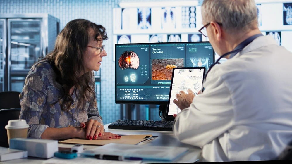
AI systems rely on image annotation services to detect diseases accurately. The process tags medical images such as X-rays, CT scans, MRI scans, mammograms, and ultrasounds. Using real-time data analysis, AI enhances the precision of medical evaluations and promotes efficient and timely diagnoses.
This piece explores how medical image annotation helps detect diseases early and improves healthcare outcomes while advancing medical research. You'll discover various annotation techniques, best practices for data preparation, and the role of quality-assured labeling in advancing medical AI applications.
How Image Labeling Supports Early Disease Detection
AI-driven diagnostic tools rely on accurate image labeling services, which form the foundation of modern healthcare priorities. Medical images with proper labels help computer systems identify disease patterns before symptoms become severe. This breakthrough in preventive healthcare allows doctors to intervene at the most effective stages of treatment.
1- Why Labeled Data is Essential for AI Diagnosis
AI diagnostic systems need labeled datasets to learn visual patterns linked to medical conditions. These supervised learning algorithms connect visual features with diagnostic conclusions. Quality medical data annotation helps create AI models that improve clinical decision-making.
AI training needs these key elements to work well:
- Balanced datasets with similar numbers of healthy and diseased subjects
- Patient data of all types to ensure algorithm generalization
- High-quality annotations that precisely identify abnormalities
- Multimodal data integration for comprehensive analysis
2- Common Use Cases: Cancer, Heart Disease, and More
Image labeling services have revolutionized the detection of life-threatening conditions. Deep learning algorithms analyze heart disease images with remarkable accuracy. For instance, a research study achieved 91.7% validation accuracy using a hybrid deep-learning model.
Cancer detection is another significant application. AI algorithms trained on labeled medical images analyze microscopic cells and identify cancer biomarkers. These algorithms detect tumors in mammogram images with 94% accuracy.
Google's AI model has shown even better results than human radiologists by reducing false positives by 5.7% and false negatives by 9.4%.
Image annotation services help detect conditions early in many medical areas:
- Neurological disorders through brain imaging analysis
- Liver disease from MRI or ultrasound images
- Dermatological conditions through skin image classification
- Pulmonary diseases via chest X-ray examinations
Preparing Medical Data for Annotation
Effective data preparation lays the groundwork for any successful AI image labeling project in medicine. You need to take several key steps before starting annotation. These steps help create datasets that can train diagnostic algorithms well.
I- Choosing the Right Image Formats (DICOM, TIFF)
The medical imaging field relies on two main formats:
DICOM (Digital Imaging and Communications in Medicine) acts as the foundation of modern medical imaging. It creates a non-proprietary way to share biomedical images. This format stores not only images but also vital metadata. These include patient information, equipment settings, and clinical details, which are important for accurate interpretation by doctors.
TIFF (Tagged Image File Format) is another common format in medical imaging. It is versatile and works with images of any size, resolution, or color depth. These files are either uncompressed or use lossless compression. This keeps the original image quality intact, though the files end up larger in size.
Your project's needs will determine the most suitable format to use. DICOM ensures consistency between different machines and manufacturers while preserving important contextual information. TIFF is best suited for scenarios that require image quality, especially for drawings and diagrams.
II- Ensuring Data Diversity and Quality
Effective annotations need diverse datasets that accurately represent reality. It’s important to collect images from different sources, equipment, and patient groups. Models trained on varied samples become more reliable compared to those trained on similar ones.
Dataset review plays a vital role too. You need to look for errors, inconsistencies, and missing data. Quality is as important as quantity. A small, high-quality dataset can match or beat larger but lower-quality ones.
III- Splitting Data for Training, Validation, and Testing
The way you split your data can make or break your model’s performance. Medical projects need splitting based on patient IDs rather than individual images. This prevents data leakage, as images from the same patient tend to look alike.
Most teams use an 80:10:10 split for training, validation, and testing. The key is to keep these splits balanced. This prevents bias that could limit your model's real-world use. Patient-based splitting gives you accurate performance metrics instead of inflated results.
Types of Medical Image Annotation and Their Applications
Medical image labeling services include a variety of annotation techniques. Each technique serves specific diagnostic needs and applications. These specialized methods help healthcare AI systems extract vital information from complex medical images.
i) Classification and Object Detection
Medical image classification assigns categorical labels to entire scans. This helps distinguish between normal and abnormal images. Object detection takes this a step further. It identifies and pinpoints specific features like tumors or lesions within images.
Object detection in medical imaging needs two key steps:
- Feature Extraction: Identifying target features within the image.
- Object Classification and Localization: Classifying and positioning the detected objects.
There are two main types of detection algorithms:
- Two-Stage Frameworks These algorithms initially extract CNN (Convolutional Neural Network) features without the category information and then apply category-specific classifiers. The common examples are R-CNN (Region-based Convolutional Neural Network) and Faster R-CNN. Two-stage frameworks are generally more accurate but slower.
- One-Stage Frameworks: These algorithms perform feature extraction and classification in one step. They are faster but generally less accurate. Examples of one-stage frameworks include SSD (Single Shot MultiBox Detector) and YOLO (You Only Look Once).
ii) Segmentation and Landmark Detection
Segmentation divides medical images into meaningful regions based on texture, brightness, and contrast. Doctors use this technique to measure tissue volumes precisely. It helps them find abnormalities and plan radiation therapy treatment.
Landmark detection identifies specific anatomical points in medical images. This helps measure distances between anatomical structures and set up surgical safety zones.
Two primary methods lead this field:
- Heatmap-Based Methods: These create probability maps where each pixel shows the likelihood of containing a landmark
- Coordinate Regression Methods: These methods directly predict the coordinates of landmarks.
iii) Semantic vs. Instance Segmentation
Semantic segmentation labels each pixel with a category, such as lung, heart, or tumor, to create detailed tissue maps. Instance segmentation works differently. It gives unique labels to separate objects of the same class. This is useful for distinguishing different tumors within a single image.
The difference between these methods is vital in medical applications. Semantic segmentation works best at understanding overall scene composition and handles immediate scenarios better due to lower computational requirements. However, it doesn't deal very well with overlapping structures.
Instance segmentation is beneficial for distinguishing individual objects of the same type. It requires more computational power but excels in scenarios with overlapping structures.
How Image Annotation Services Drive Medical Research
Quality-annotated medical images are pivotal in advancing healthcare research. Medical data contains essential patterns and insights that image labeling services help reveal.
1) Supporting Algorithm Development and Validation
Labeled datasets are vital building blocks for reliable AI algorithms in medical imaging. The annotation process must ensure accuracy, consistency, and completeness to make algorithms work correctly. Without meeting these quality standards, AI systems can produce unreliable results.
Annotated datasets are important for obtaining accurate results during validation. Many AI systems fail to work in clinical settings because they lack external dataset validation.
Validation datasets created through pathologist annotations allow developers to thoroughly test algorithm performance before clinical use.
2) Standardizing Data for Multi-Center Studies
Standardization improves research quality by improving integration and data reuse. Researchers need standardization to combine data from multiple sources while ensuring consistency throughout.
Standardization helps researchers ‘speak the same language’ by aligning vocabularies and data formats. This shared framework speeds up recruitment, covers diverse populations, and makes multi-center research more relevant.
Enhancing Medical Education with Annotated Datasets
Annotated medical image datasets serve as powerful teaching tools beyond their clinical applications.
3) Educational datasets serve three main purposes:
- Medical students learn to identify normal and pathological patterns
- Researchers can test new analytical approaches
- Teams can compare algorithmic performance against established standards
Educational datasets must include diverse patient populations to avoid algorithmic bias.
Image labeling outsourcing services provide specialized educational resources that help overcome the challenge of accessing well-annotated datasets from different ethnicities.
Conclusion
Medical image annotation services are important for modern healthcare progress. These services help doctors detect diseases with precision, support groundbreaking research, and boost medical education through well-labeled datasets.
High-quality image labeling facilitates earlier diagnosis and more accurate treatment planning. Standardized data preparation and annotation techniques provide a solid foundation for healthcare breakthroughs.
Medical teams achieve better results in classification, segmentation, and landmark detection. This makes diagnostic processes faster and treatments more accurate.
The future of healthcare delivery and research heavily relies on medical image annotation. Healthcare providers who adopt these services can deliver superior patient care and advance medical knowledge for future generations.


Sign in to leave a comment.