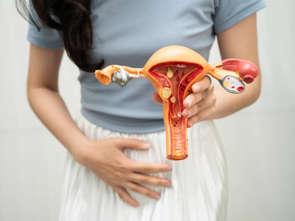


Understanding Calcified Uterine Leiomyoma
A calcified uterine leiomyoma is a type of uterine fibroid that has developed areas of calcium deposits within the fibroid tissue. Uterine leiomyomas are noncancerous growths that develop from the muscular tissue of the uterus. Over time, some fibroids may undergo changes caused by reduced blood supply, degeneration, aging, or other tissue changes. Calcium can gradually accumulate in these areas, creating what is commonly described as a calcified uterine leiomyoma.
Not every calcified fibroid causes noticeable symptoms. Some are discovered during routine pelvic imaging when a person is being evaluated for another concern. Others may contribute to pelvic pressure, discomfort, heavy menstrual bleeding, abdominal fullness, urinary symptoms, or changes in bowel habits. The symptoms often depend on the size, location, and number of fibroids rather than calcification alone.
Understanding the nature of a calcified uterine leiomyoma is important because treatment decisions should be based on the overall clinical picture. A healthcare professional may consider symptoms, imaging findings, fibroid location, reproductive goals, general health, and previous treatments before recommending an appropriate approach.
How Calcification Develops
Fibroids can change over time as their blood supply and internal structure change. When part of a fibroid receives less blood flow, the tissue may undergo degeneration. In some cases, calcium deposits develop in the affected tissue. This process can make the fibroid appear harder or more densely calcified on imaging.
Calcification does not automatically mean that a fibroid is dangerous. A calcified uterine leiomyoma is generally associated with a benign fibroid process. However, the presence of calcification does not by itself determine whether treatment is necessary. A healthcare provider needs to evaluate the entire imaging appearance and the patient's symptoms.
Imaging studies such as ultrasound, magnetic resonance imaging, or computed tomography may help identify the size and position of a fibroid and determine whether calcification is present. These findings can help guide the next steps and distinguish a stable fibroid from conditions that may require additional evaluation.
When Treatment May Be Considered
Treatment is not always necessary for a calcified uterine leiomyoma. If the fibroid is small, stable, and not causing significant symptoms, a healthcare provider may recommend observation and periodic follow-up. Monitoring can help determine whether the fibroid changes over time.
Treatment may become appropriate when symptoms interfere with daily activities or quality of life. Heavy or prolonged menstrual bleeding can be particularly disruptive and may contribute to tiredness or anemia. Pelvic pressure and discomfort may also affect work, exercise, sleep, and normal routines.
A calcified uterine leiomyoma may also require treatment consideration when it becomes large enough to place pressure on nearby organs. Depending on its location, a fibroid can contribute to frequent urination, difficulty emptying the bladder, constipation, or a sensation of abdominal fullness.
Medication-Based Management
Medications may be used to manage symptoms associated with uterine fibroids. Depending on the individual's symptoms and medical history, healthcare providers may recommend medications that help reduce heavy menstrual bleeding or relieve pelvic discomfort.
Medication generally focuses on symptom control rather than removing the fibroid itself. Therefore, it may be more useful for people whose primary concern is bleeding or pain and who do not require a procedure immediately.
For someone with a calcified uterine leiomyoma, medication decisions should be individualized. The presence of calcification may be only one part of the overall assessment. A healthcare professional can determine whether medication is appropriate based on symptoms, imaging, age, reproductive plans, and other health considerations.
Uterine Fibroid Embolization
Uterine fibroid embolization is a minimally invasive treatment option for selected patients with symptomatic fibroids. During the procedure, a specialist uses image guidance to access the blood vessels supplying the fibroid. Small particles are then introduced to reduce blood flow to the targeted fibroid.
As the blood supply decreases, the fibroid can shrink over time and symptoms may improve. Uterine fibroid embolization is designed to treat the fibroid without requiring traditional open surgery.
For patients with a calcified uterine leiomyoma, treatment planning requires careful imaging evaluation. Calcification can alter the internal characteristics of a fibroid, so the treating specialist needs to understand the fibroid's location, blood supply, and relationship with surrounding structures before deciding whether embolization is suitable.
Image-Guided Treatment Planning
Imaging plays an important role in the evaluation of a calcified uterine leiomyoma. Ultrasound can provide information about the uterus and fibroids, while MRI can offer detailed information about fibroid size, location, tissue characteristics, and surrounding anatomy.
Detailed imaging may help a specialist determine whether symptoms are likely to be related to a particular fibroid. It can also assist in selecting a treatment approach that fits the patient's individual circumstances.
Image-guided procedures can provide a less invasive alternative to traditional surgical approaches for appropriately selected patients. However, not every fibroid or every patient is suitable for the same treatment. A personalized evaluation remains essential.
Surgical Treatment Options
Surgery may be considered when fibroids are causing significant symptoms, are very large, have a particular location, or have not responded adequately to other treatment approaches. The type of surgery depends on the patient's circumstances and treatment goals.
Myomectomy is a surgical procedure that removes fibroids while preserving the uterus. It may be considered for patients who want to retain their uterus, including some people who are planning future pregnancy. The surgical approach can vary depending on fibroid size, number, and location.
Hysterectomy involves removing the uterus and provides a definitive treatment for uterine fibroids because fibroids cannot grow in a uterus that has been removed. However, it is a major decision and is not appropriate for everyone. A healthcare provider can explain the potential benefits, risks, recovery considerations, and alternatives.
When a fibroid has substantial calcification, the surgeon may consider the fibroid's firmness and location when planning the procedure. Imaging can provide useful information before surgery and help the medical team understand the anatomy involved.
Observation and Follow-Up
For some people, monitoring a calcified uterine leiomyoma may be the most appropriate approach. Observation can be particularly reasonable when symptoms are minimal and imaging does not show concerning features.
Follow-up may involve periodic clinical assessments and imaging when recommended by a healthcare professional. Monitoring allows changes in symptoms or fibroid characteristics to be identified over time.
Patients should communicate any new or worsening symptoms to their healthcare provider. Increasing pelvic pain, significant changes in menstrual bleeding, pressure symptoms, or other new concerns may warrant another evaluation.
Managing Heavy Menstrual Bleeding
Heavy menstrual bleeding is one of the common concerns associated with uterine fibroids. When bleeding becomes excessive, it can affect daily life and may contribute to iron deficiency or anemia.
Treatment depends on the cause and severity of the bleeding. A healthcare provider may recommend medication, a minimally invasive procedure, or surgery depending on the fibroid characteristics and the patient's overall needs.
A calcified uterine leiomyoma may not always be the direct cause of bleeding, so evaluation should consider other possible causes as well. Identifying the source of symptoms is an important part of choosing an effective treatment strategy.
Managing Pelvic Pressure and Discomfort
Fibroids can produce a feeling of pressure or fullness in the lower abdomen or pelvis. Larger fibroids may press against surrounding structures and contribute to urinary or bowel symptoms.
Pain can have several possible causes, including fibroid degeneration, pressure on nearby tissues, or other gynecological conditions. Calcification itself does not necessarily mean that a fibroid will cause pain.
When discomfort persists, an appropriate medical evaluation can help determine whether the fibroid is responsible and whether treatment could provide meaningful relief.
Choosing the Right Treatment
Choosing treatment for a calcified uterine leiomyoma requires more than simply looking at whether calcification is present. The size, location, number, symptoms, imaging appearance, and overall health of the patient all matter.
Personal priorities are also important. Someone who wants to preserve the uterus may consider different options from someone who has completed childbearing and wants a definitive treatment. Recovery preferences, previous procedures, and individual medical circumstances can also influence the decision.
A specialist can discuss available approaches and explain which options may be appropriate. Patients should feel comfortable asking about expected outcomes, recovery, potential complications, and alternatives before making a treatment decision.
The Importance of Personalized Care
Every fibroid case is different. Two people may both have a calcified uterine leiomyoma but experience completely different symptoms and require different management strategies. One person may need only observation, while another may benefit from an intervention because of persistent bleeding, pressure, pain, or other symptoms.
Personalized care allows treatment to focus on the patient's specific needs rather than relying on a single approach for everyone. A thorough evaluation can help determine whether minimally invasive treatment, medication, surgery, or continued monitoring is most appropriate.
What Patients Should Discuss With a Specialist
During an appointment, patients can ask about the size and location of their fibroid, the significance of calcification, and whether the fibroid is likely responsible for their symptoms. It is also helpful to ask what treatment options are available and what benefits and limitations each approach may have.
Questions about recovery, future pregnancy plans, preservation of the uterus, symptom improvement, and the possibility of additional treatment can also help patients make informed decisions.
Understanding the condition and available choices can make the treatment process less stressful. A specialist can review imaging and medical history and explain the options in language that is easy to understand.
Final Thoughts
A calcified uterine leiomyoma is a fibroid that has developed calcium deposits within its tissue, often as part of changes that occur over time. Although calcification can make a fibroid appear different on imaging, it does not automatically mean that treatment is required.
Management may include observation, medication for symptom control, minimally invasive procedures such as uterine fibroid embolization, or surgical treatment such as myomectomy or hysterectomy. The best approach depends on symptoms, fibroid characteristics, imaging findings, reproductive goals, and overall health.
If a calcified uterine leiomyoma is causing persistent bleeding, pelvic pressure, pain, or other concerns, discussing the condition with a qualified specialist can help clarify the available options. With appropriate evaluation and individualized care, patients can better understand their condition and choose a treatment approach that supports their health and quality of life.


















Sign in to leave a comment.