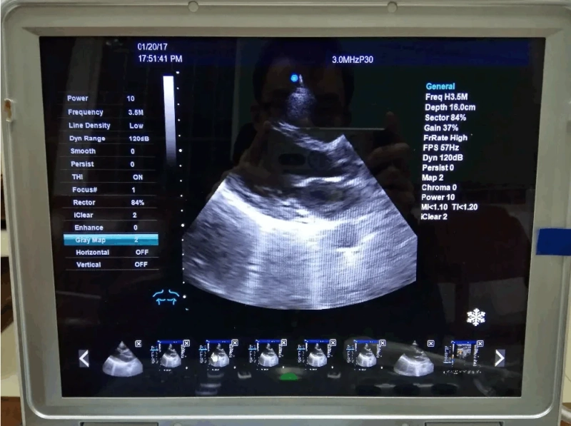
Ultrasound color Doppler, also known as color Doppler, is a medical device that is suitable for ultrasound examination of various parts of the body, especially for the examination and diagnosis of the heart, limb blood vessels and superficial organs, as well as the abdomen, obstetrics and gynecology.
Features
Its working principle is based on the introduction of color Doppler technology on the basis of high-definition black and white B ultrasound . Color Doppler ultrasound blood flow images can be formed, which not only has the advantages of two-dimensional ultrasound structural images, but also provides rich information on hemodynamics. It is clinically known as "non-invasive angiography ".
color identification
Color Doppler blood flow imaging technology is to display blood flow signals in color, and the pseudo-color coding is composed of three basic colors of red, blue and green. Set red to represent blood flow towards the probe and blue to represent blood flow away from the probe. The blood flow velocity is related to the color brightness, high speed, strong color brightness, low speed, weak color brightness. For example, when the blood flow velocity toward the probe is low, the signal is dark red, and when the blood flow velocity away from the probe is low, the signal is dark blue. Difficulty distinguishing on the screen. At this time, a color intensifier is added to improve the color signal brightness of low-velocity blood flow. In order to express the blood flow rate accurately and quickly, sometimes three colors are used to indicate the speed of blood flow, and the signal from dark red to bright red is used to indicate the blood flow toward the probe. If the blood flow rate is faster, it will change from red to yellow. , the yellow turns green, and the coexistence of the three colors indicates different flow rates. Faster flow rates of blood flow away from the probe are indicated in cyan, green.
Two color maps are set on the ultrasound instrument: one is used for blood flow detection of non-cardiovascular system, and only has two colors of red, yellow, blue and blue; the other is used for blood flow of cardiovascular system. Flow detection has two to three colors in each direction.
The main difference with B-ultrasound:
1. B-ultrasound has only one probe, which can only examine abdominal organs in general; while color has three probes, which can examine the heart, superficial skin, blood vessels, tumors, etc. in addition to the abdominal cavity.
2. The main technical indicators of color ultrasound are much higher than those of B ultrasound (such as the number of probe elements, the number of imaging channels, the imaging dynamic paradigm, the processing capacity and speed of the host, etc.) Smaller lesions are found, improving the early diagnosis rate of the disease, and can more clearly show the details of changes around and within the lesion.
3. Color Doppler has the function of color Doppler blood flow imaging, which can display the changes of vascular anatomy, blood flow direction, blood flow velocity and blood flow state in the lesion area, which can significantly improve the ability of differential diagnosis of diseases and improve the accuracy of diagnosis. sex.
4. Color Doppler has the function of tissue harmonic imaging, which can significantly reduce the interference of obesity, gas and other artifacts, and improve the clarity of the image.
5. Color Doppler ultrasound has the function of contrast agent harmonic imaging, which can perform contrast-enhanced ultrasound to conduct more in-depth examination and research on lesions.
Clinical application
1. Vascular disease
Using a 10MHz high-frequency probe can find calcification points less than 1mm in the blood vessel, which has a good diagnostic value for carotid atherosclerotic occlusive disease. It can also use the local magnification of blood flow exploration to determine the degree of lumen stenosis, whether the emboli have fallen off, and whether Ulcers were formed, preventing the occurrence of cerebral embolism.
It is the best diagnostic method for various types of arteriovenous fistulas, and the diagnosis can be made when the colorful mosaic ring spectrum is detected.
For carotid aneurysm, abdominal aortic aneurysm, vasculitis obliterans, chronic lower extremity veins and other diseases, the use of high-definition color Doppler ultrasound, local magnification and blood flow spectrum exploration can make a more accurate diagnosis.
2. Abdominal organs
Mainly used for liver and kidney diagnosis. It is an auxiliary diagnosis for the identification of benign and malignant lesions in the abdominal cavity, the identification of gallbladder cancer from large polyps, chronic severe inflammation, the difference between common bile duct and hepatic artery and other diseases.
3. Small Organs
Among the small organs, the main ones are the thyroid , breast , eyeball , etc.
4. Prostate and seminal vesicles
With prostate biopsy, various prostate seminal vesicle gland diseases can be basically diagnosed.
5. Obstetrics and Gynecology
For the identification of benign and malignant tumors and umbilical cord disease, fetal congenital heart disease and assessment of placental function. It can make a better diagnosis of infertility and pelvic varicose veins, and has better auxiliary diagnostic value for trophoblastic diseases. The use of vaginal probes has certain advantages over abdominal exploration, which are mainly reflected in the following aspects:
1) It is sensitive to uterine artery and ovarian blood flow and has a high display rate.
2) Shorten inspection time and obtain accurate Doppler spectrum.
3) No need to fill the bladder.
4) Does not interfere with body type obesity, abdominal scars, and intestinal inflation.
5) Use the movement of the probe tip to find the tender part of the pelvic organs to determine whether there is adhesion in the pelvis.
Classification
According to their clinical use, color Doppler ultrasound diagnostic instruments can be divided into the following types:
1. Abdominal special type: The basic functions include two-dimensional ultrasound, color Doppler blood flow imaging, color power Doppler, pulse wave spectrum Doppler and other technologies.
2. Cardiovascular-specific type: The main function is the same as the abdominal type, but with the addition of continuous wave spectral Doppler, deletion of color power Doppler, addition of M-mode ultrasound, or with Doppler tissue imaging (TDL) and other functions.
3. Dual-purpose or universal type: It has the functions of abdominal and cardiovascular color Doppler
Precautions
1. The digestive system examination requires the patient to fast for more than 8 hours.
2. Urinary system and obstetrics and gynecology disease examination requires the patient to fill the bladder.
3. Those who do gastrointestinal angiography are required to do ultrasonography after three days.
advantage
1. Quickly and intuitively display the two-dimensional plane distribution of blood flow.
2. Displays the direction of blood flow.
3. Facilitates the identification of arteries and veins.
4. Identify vascular and non-vascular lesions.
5. Understand the nature, direction and speed of blood flow.
6. Quantitative analysis of the origin, width, length and area of blood flow.
Environmental requirements
The higher the equipment, the higher the environmental requirements, including temperature, humidity, dust, etc.
1. The best indoor temperature is 22-27℃, and the preheating should be turned on first in winter.
2. The humidity should be maintained at 40% to 60%. The humidity is high in summer and the yellow plum season. The ultrasonic equipment should be placed on the second floor or above. In addition to ensuring the dehumidification of the air conditioner, a dehumidifier should be equipped when necessary to avoid high humidity corrosion of circuit components and shorten the life of the instrument.
3. The ultrasound department should be far away from equipment with strong electromagnetic fields such as X-ray machines, CT machines, and high-frequency treatment machines to ensure that the instruments are not disturbed.
4. Ultrasound equipment should be avoided to be installed beside noisy roads with heavy traffic flow to prevent noise interference and a lot of dust. The staff asked to enter the studio to change shoes, and the patient also asked to wear shoe covers.
5. Ultrasound equipment should also be avoided to be placed near Chinese and Western pharmacies, canteens, etc. The doors and windows should be tight and seamless, and food should not be placed indoors to prevent mice from gnawing on the probe and cables
Requirements
1. The power supply should be equipped with a dedicated line circuit and an electronic AC voltage stabilizer with good performance, high voltage stabilization accuracy, interference purification function, and overvoltage protection function. The power of the voltage stabilizer should be 2 to 3 times greater than the power of the instrument.
2. The grounding resistance of the ground wire should be less than 4 ohms. Good grounding can reduce the interference of external electrical interference signals to the equipment.
3. The instrument should be used by a dedicated person. After daily use, clean the probe and remove the couplant on the table to prevent the couplant from sticking to the buttons on the table and flowing into the instrument and corroding the electronic board.
4. Regularly clean and maintain the appearance of the instrument. Clean the filter every 1 to 2 weeks
Analysis and response to common faults
The most common failure is "crash". The instrument will "crash" when it is "too hot", and the interference such as transient current will also "crash". These situations can be eliminated by cooling down, clearing the filter, and powering off. First, disconnect the power supply at the first time, consider external influences, exclude external circuit interference, and check whether the temperature and humidity are suitable. Secondly, pay attention to the instrument itself, check whether the dust removal net is cleaned or not, after 10 minutes after disconnecting the power supply, turn on the voltage stabilizer first, wait for the voltage to stabilize before turning it on again, and observe whether there are any abnormal signals in each step of the startup, so as to analyze the "crash" "What's the problem. If there are obvious software and hardware failures during the self-inspection process, it must be reported to a professional engineer for repair.
https://www.arshinemedical.com/news/what-is-a-color-doppler-ultrasound-system-60454991.html


