

Automated hematology analyzers can quickly investigate whole blood examples for the complete blood count (CBC). Outcomes comprise red blood cell (RBC) count, white blood cell (WBC) count, platelet count, hemoglobin absorption, hematocrit, RBC guides, and a leukocyte discrepancy. Less hi-tech automated hematology analyzers supplied by Hematology Analyzer Suppliers in a physician’s office location may occasionally offer a limited CBC, using older technology for whole blood examination (e.g., impedance technology) that will produce only a three-part leukocyte differential. A three-part leukocyte disparity delivers values for neutrophils, lymphocytes, and all other white cells together. More modern Automated hematology analyzers are proficient in examining all leukocytes consuming flow cytometry-based approaches, some in grouping with cytochemistry or fluorescence or conductivity, to count all the diverse kinds of WBCs, including neutrophils, lymphocytes, monocytes, basophils, and eosinophils (five-part differential). Nucleated RBCs are also noticed.
Leukocytosis or raised WBC count is a shared laboratory finding. For instance, a WBC count of 30×109/L (30,000/μL) is irregular in an adult but normal in a newborn within the first few days of life. The usual WBC differential also modifies with age, and proper usual ranges must be recognized for each laboratory carrying out such tests. Leukocytosis is also a highlight of leukemias; thus, singularizing leukemias from other sources of leukocytosis is vital. Inspection of outlying blood smear along with an appraisal of CBC examination is vital for such differentiation, and if essential, further examination such as flow cytometry, molecular studies, and possible bone marrow inspection must be commenced. Correspondingly, platelet complaints such as thrombocytopenia or thrombocytosis may be a non-threatening disorder or may indicate a serious disorder such as severe thrombocytopenia observed in patients with acute leukemias.
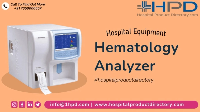
Examination of Various Parameters by Automated hematology analyzers:
Modern Automated hematology analyzers are capable of counting as well as defining the size of numerous mingling blood cells in the blood, including RBCs, WBCs, and platelets. One such tool, the Beckman–Coulter analyzer, produces an electrical pulse when a blood cell passes through the analyzer station, which comprises a small opening enclosed by electrodes. Each electrical pulse signifies an individualistic cell, and pulse height designates the cell volume. Modern Automated hematology analyzers are also capable of multimodal valuation of cell size and cell count, thus providing supplementary information concerning various groups of WBCs, such as neutrophils, lymphocytes, monocytes, eosinophils, and basophils.
Optical Fluorescence Platelet Counting:
An optical fluorescence platelet total is also done on Sysmex XE-2100 and XE-5000 analyzers, in addition to the old-style impedance sum. The optical fluorescent platelet count is calculated in the reticulocyte station. A polymethine color is used to tint the RNA/DNA of reticulated cells and platelet skins and granules. This skill permits the concurrent counting of the red cell reticulocytes, erythrocytes, and fluorescent platelets. Within the stream cell, every single cell is passed through the light ray of a semiconductor diode laser. The fluorescence strength of each cell is examined, which permits the separation of platelets from red cells and reticulocytes. The fluorescent discoloration of the platelets not only permits the exclusion of nonplatelet particles from the count but also permits the addition of large or giant platelets.
Mistakes in Platelet Count:
In certain circumstances, Automated hematology analyzers supplied by Automated hematology analyzers in India are recognized to deliver a falsely low platelet count when the true platelet count is satisfactory. This may give growth to suboptimal clinical administration. Incomplete clotting for example or platelet activation during venipuncture may reason platelet aggregation. Both methods may steer to low platelet counts. Confirming the example for clots, examining the histograms, as well as studying the smear are all significant steps to evade misleading low platelet counts. There are numerous other methods to clarify falsely low platelet counts,
Otherwise mentioned as pseudo thrombocytopenia: anticoagulant-induced pseudo thrombocytopenia, platelet satellites, giant platelets, and cold agglutinin-induced platelet agglutination. Incorrectly raised platelet counts are much less shared than falsely low counts. Disjointed red cells or white cell remains may be totaled as platelets, giving rise to high platelet counts. Disjointed red cells can be seen in conditions of microangiopathic hemolysis such as dispersed intravascular coagulation (DIC). White cell wreckages can be understood in leukemic or lymphoma states. Patients with leukemia, particularly acute leukemia, require supportive treatment in the form of blood component transfusions. If the platelet count is incorrectly raised in a patient with acute leukemia, the choice to transfuse platelets may be deferred, with unwanted clinical significance. Incorrectly high platelet counts may also be understood in the attendance of cryoglobulins and microorganisms present in the blood.
0