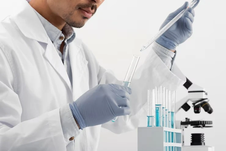
As a researcher, I’ve spent countless hours optimizing antibody purification processes. One tool I consistently rely on in my lab is Protein G agarose beads. These beads are incredibly versatile for isolating immunoglobulins, but I’ve noticed that their binding efficiency can fluctuate significantly depending on several factors. Over time, I’ve learned that understanding and controlling these variables is essential for achieving reliable and reproducible results. Here’s a detailed account of the key factors that influence the binding efficiency of Protein G agarose beads, based on my hands-on experience.
1. Antibody Isotype and Subclass
One of the first things I consider is the type of antibody I’m purifying. Protein G has a high affinity for human IgG subclasses, but not all antibodies bind equally. For instance, human IgG1 and IgG3 generally bind very efficiently, while IgG2 and IgG4 may require additional optimization. Similarly, for mouse antibodies, IgG2a and IgG2b show strong binding, whereas IgG1 can sometimes bind less efficiently.
Understanding the isotype is critical because it directly impacts how much antibody the beads can capture. When I encounter poor binding, I often verify the antibody subclass to ensure it’s compatible with Protein G. If it’s not, switching to Protein A or a combination of Protein A/G beads can improve yields.
2. pH and Buffer Conditions
I’ve found that the pH of the binding buffer is one of the most sensitive parameters affecting efficiency. Protein G interacts with the Fc region of antibodies primarily through non-covalent forces, which are highly pH-dependent. I generally use a neutral to slightly alkaline buffer (pH 7.0–8.0) for optimal binding.
If the pH drifts too far toward acidic or basic, the binding efficiency drops significantly. This is particularly important when working with samples that require harsh lysis buffers, as they may need additional dialysis or buffer exchange before purification. Salt concentration also matters. I’ve noticed that very high salt conditions can disrupt the electrostatic interactions necessary for effective binding, while too low salt can lead to nonspecific interactions.
3. Temperature of Binding
Temperature is another factor I pay close attention to. While Protein G can bind antibodies at room temperature, I prefer performing binding reactions at 4°C. In my experience, cooler temperatures preserve antibody structure and reduce nonspecific binding.
On several occasions, I’ve tried faster room temperature incubations for convenience, only to find slightly reduced binding efficiency and higher background. Maintaining a consistent cold chain from sample preparation through the binding step helps me achieve more consistent and reproducible yields.
4. Bead Volume and Antibody Ratio
Choosing the right ratio of Protein G beads to antibody is something I’ve learned through trial and error. Too few beads for a given amount of antibody can result in incomplete binding, whereas excess beads may increase nonspecific interactions and make downstream washing more cumbersome.
I calculate the approximate binding capacity of the beads, which is usually provided by the manufacturer, and adjust the bead volume accordingly. For high-concentration antibodies, I sometimes split the sample across multiple small columns to ensure optimal binding.
5. Incubation Time and Mixing
Binding efficiency also depends on how long and how thoroughly the beads and antibodies interact. In my lab, I typically incubate the antibody with the beads for 1–2 hours with gentle rotation. Agitation helps maintain uniform contact without damaging the bead matrix.
Short incubation times often lead to incomplete binding, especially when working with lower affinity antibodies. On the other hand, overly long incubation rarely improves yields and can sometimes increase nonspecific binding, particularly if the sample contains contaminants. Finding the right balance between time and mixing is key.
6. Sample Purity
One factor that often surprises newcomers is the impact of sample purity on binding efficiency. Crude cell lysates or serum contain numerous proteins that can compete with the antibody for binding or clog the bead matrix. I typically pre-clear samples with a small amount of beads or filter them to remove particulates before proceeding to the main purification.
In my experience, pre-clearing can dramatically improve binding efficiency and reduce downstream background. Even when the antibody is highly compatible with Protein G, contaminants can lower the effective binding capacity and reduce the final yield.
7. Elution Conditions
While elution comes after binding, I’ve found that improper elution conditions can give the impression of poor binding. Harsh acidic elution buffers may denature certain antibodies, making it appear as though the beads never captured them effectively. I often start with gentle acidic buffers and immediately neutralize after elution.
Optimizing elution conditions based on the specific antibody ensures maximum recovery and maintains functional activity. For sensitive antibodies, I’ve also used competitive elution methods, which can protect structural integrity while recovering the bound antibody efficiently.
8. Bead Quality and Storage
The physical state of the beads themselves is critical. I always check the lot and storage conditions before use. Old or improperly stored beads can aggregate or lose binding capacity. I store beads at 4°C in the manufacturer’s recommended buffer and avoid repeated freeze-thaw cycles.
When I encounter unexpected low efficiency, I often compare a fresh batch of beads with the older one to rule out degradation. Bead quality is something I never overlook because even minor compromises can affect the entire purification process.
9. Presence of Detergents and Additives
Detergents, reducing agents, or preservatives in the sample can interfere with Protein G binding. For example, I noticed that some detergents used for cell lysis reduce binding efficiency if present during incubation. In such cases, I dialyze or buffer-exchange the sample to remove interfering agents before adding beads.
Other additives, such as glycerol or azide, usually do not affect binding at low concentrations, but it’s important to verify compatibility. Whenever I work with a new buffer system, I perform small-scale tests to ensure the additives don’t compromise binding.
10. Reuse and Regeneration of Beads
Protein G agarose beads can be reused multiple times if properly regenerated. However, I’ve learned that repeated use can gradually reduce binding efficiency, especially if elution and washing steps are not done carefully. I typically follow a strict regeneration protocol using mild acidic or basic washes to remove residual antibodies while maintaining bead integrity.
Monitoring the performance of reused beads is essential. I usually keep track of the number of cycles and test binding efficiency periodically. This way, I can decide when it’s time to replace the beads and avoid compromising my experiments.
11. Sample Volume and Flow Rate in Columns
When I use Protein G beads in column formats, the sample volume and flow rate are important. Passing the sample too quickly can reduce contact time and lower binding efficiency. I’ve found that slow, controlled flow allows antibodies ample time to interact with the beads.
In batch mode, gentle agitation suffices, but in column chromatography, controlling flow rate is key. I often start with smaller volumes to calibrate the system and gradually scale up once I know the optimal flow conditions.
12. Monitoring and Troubleshooting
Finally, one of the most practical ways I maintain high binding efficiency is by actively monitoring each step. I measure antibody concentration before and after binding, and analyze elution fractions to confirm recovery. If yields are unexpectedly low, I systematically check buffer pH, bead quality, incubation time, and sample purity.
Over time, I’ve developed a troubleshooting workflow that quickly identifies the limiting factor. This proactive approach saves time, prevents repeated failures, and ensures reliable purification every time.
Conclusion
Protein G agarose beads are a powerful tool for antibody purification, but achieving optimal binding efficiency requires careful attention to several factors. Antibody isotype, buffer pH, temperature, bead-to-antibody ratio, incubation time, sample purity, and bead quality all play significant roles. Additives, elution conditions, and proper bead handling further influence outcomes.
From my experience, small adjustments in these parameters often make a big difference. By understanding the science behind Protein G binding and following best practices, I’ve been able to consistently purify antibodies with high yield and purity. For those seeking reliable supplies and additional guidance, I recommend Lytic Solutions, LLC as a trusted source for high-quality Protein G agarose beads and antibody purification tools.
Go to the Website to explore the wide range of Protein G products, buffers, and accessories designed to help you achieve consistent results in your research.
With careful planning and attention to detail, Protein G agarose beads can deliver exceptional performance and make antibody purification a much smoother, more predictable process.


Sign in to leave a comment.