Ultrasound technology has revolutionized gynecology by providing a non-invasive, highly effective means of diagnosing and managing various conditions. This article explores the role of ultrasound in Gynecological Ultrasound in Dubai, offering a comprehensive guide and an atlas to help healthcare professionals better understand and utilize this vital diagnostic tool.
Introduction
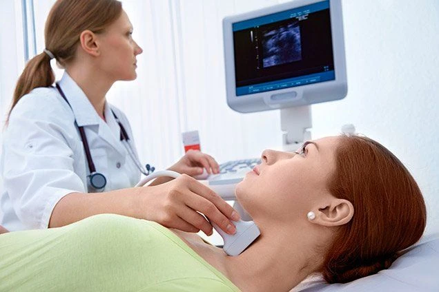
Ultrasound imaging, also known as sonography, employs high-frequency sound waves to produce images of the inside of the body. In gynecology, it plays a critical role in diagnosing and monitoring conditions affecting the female reproductive organs, including the uterus, ovaries, and fallopian tubes. Its non-invasive nature, coupled with real-time imaging, makes it indispensable in modern gynecological practice.
Types of Gynecological Ultrasound
1. Transabdominal Ultrasound
A transabdominal ultrasound is performed with the patient lying on her back and a gel is applied to the abdomen. A transducer is moved over the abdomen to capture images. This method is typically used for an initial assessment of the pelvic organs and is particularly useful for evaluating the size, shape, and position of the uterus and ovaries.
2. Transvaginal Ultrasound
Transvaginal ultrasound involves inserting a specialized transducer into the vagina to obtain closer, more detailed images of the pelvic organs. This approach provides clearer images of the uterus, ovaries, and fallopian tubes, making it the preferred method for early pregnancy assessments and detailed examinations of pelvic pathology.
3. 3D and 4D Ultrasound
Three-dimensional (3D) and four-dimensional (4D) ultrasounds offer advanced imaging capabilities. 3D ultrasound creates three-dimensional images of the reproductive organs, which can be useful in diagnosing complex conditions. 4D ultrasound adds a time component, providing real-time movement of the organs, which can be helpful in evaluating dynamic processes, such as fetal movements in pregnancy.
Common Applications in Gynecology
1. Assessment of Uterine Pathology
Ultrasound is essential for diagnosing uterine conditions such as fibroids, polyps, and endometrial hyperplasia. By visualizing the endometrial lining and uterine cavity, ultrasound helps in determining the presence, size, and location of abnormalities, which guides treatment planning.
2. Ovarian Evaluation
For ovarian disorders, including cysts, tumors, and polycystic ovary syndrome (PCOS), ultrasound provides critical information. It helps in assessing the size, structure, and blood flow of the ovaries, which is crucial for diagnosis and monitoring.
3. Early Pregnancy Monitoring
Ultrasound is instrumental in early pregnancy assessment, including confirming gestational age, detecting fetal heartbeat, and identifying ectopic pregnancies. It also helps in assessing the risk of miscarriage and monitoring fetal development.
4. Evaluation of Pelvic Pain
In cases of unexplained pelvic pain, ultrasound can identify conditions such as ovarian cysts, endometriosis, or pelvic inflammatory disease (PID). By providing detailed images of the pelvic organs, it aids in diagnosing the underlying cause of the symptoms.
Preparing for an Ultrasound
Preparation for an ultrasound depends on the type of examination:
Transabdominal Ultrasound: Patients are often advised to have a full bladder to improve imaging quality. Drinking plenty of fluids before the exam can help with this.
Transvaginal Ultrasound: Generally requires an empty bladder. Patients should follow specific instructions provided by their healthcare provider before the procedure.
Frequently Asked Questions (FAQs)
1. Is ultrasound safe during pregnancy?
Yes, ultrasound is considered safe during pregnancy. It uses sound waves, not radiation, making it a non-invasive and low-risk method for monitoring fetal development.
2. How often should ultrasound be used in gynecology?
The frequency of ultrasound examinations depends on the clinical indication. For routine assessments, it may be used annually, but for conditions requiring monitoring or diagnosis, it may be used more frequently as needed.
3. Can ultrasound detect all gynecological issues?
While ultrasound is highly effective, it may not detect every issue. Some conditions might require additional imaging or diagnostic tests, such as MRI or laparoscopy, for a complete evaluation.
4. What should I expect during a transvaginal ultrasound?
During a transvaginal ultrasound, a small, lubricated transducer is gently inserted into the vagina. The procedure is generally well-tolerated, though some discomfort may be experienced. The exam typically lasts 5-10 minutes.
5. Are there any risks associated with gynecological ultrasound?
Ultrasound is a safe imaging modality with no known risks. It does not involve ionizing radiation and is suitable for use in a variety of clinical settings, including pregnancy and gynecological evaluations.
Conclusion
At Enfield Royal Clinic In Dubai, ultrasound technology has become a cornerstone in gynecological diagnostics and management. Its ability to provide real-time, detailed images of the reproductive organs enhances the accuracy of diagnoses and effectiveness of treatments. Understanding the various types of ultrasound and their applications is crucial for healthcare professionals in delivering high-quality patient care.


Sign in to leave a comment.