

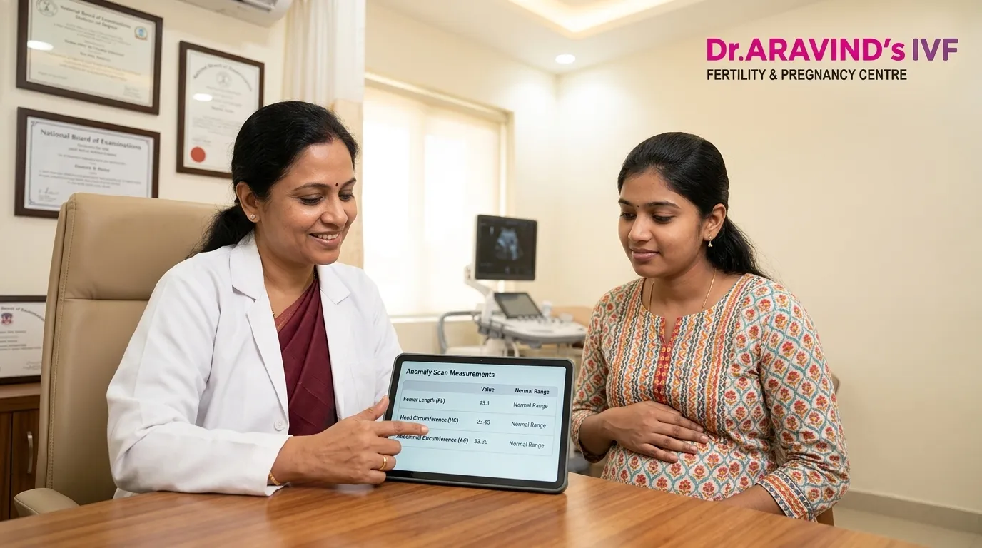
The anomaly scan appointment sits at the midpoint of pregnancy — and carries a significance that no other routine scan quite matches. It is the most comprehensive structural assessment of the developing baby available before birth — and understanding what it measures, what normal looks like across every system, and what the results genuinely mean gives every pregnant woman the confidence to face this appointment prepared rather than anxious. At Dr. Aravind's IVF Fertility and Pregnancy Center, home of a trusted IVF doctor in Karur, we believe complete information before the scan consistently produces better emotional outcomes than reassurance after it.
What Is the Anomaly Scan?
The Mid-Pregnancy Assessment That Looks at Everything
The anomaly scan — also called the mid-trimester ultrasound, level 2 scan, or TIFFA scan (Targeted Imaging for Fetal Anomalies) — is performed between 18 and 22 weeks of pregnancy, with 20 weeks considered the optimal timing for the most complete structural assessment.
Unlike earlier scans that primarily confirm viability and dating, the anomaly scan systematically examines every major organ system and structural component of the developing baby — providing the most detailed picture of fetal health available through ultrasound technology.
What the Anomaly Scan Examines
The Complete Structural Assessment
Brain and central nervous system:
The cerebral ventricles, cerebellum, corpus callosum, and posterior fossa are each assessed for normal development and fluid levels.
Face and profile:
Lip continuity, nasal bone presence, and facial profile are examined for structural normality.
Heart — four-chamber view and outflow tracts:
The most detailed cardiac assessment available by routine ultrasound — examining chamber size, septal integrity, valve function, and great vessel positioning.
Spine:
The entire spine is assessed in longitudinal and transverse views for neural tube defects and vertebral integrity.
Abdominal organs:
Stomach, kidneys, bladder, liver, and bowel are each examined for normal appearance and position.
Limbs:
All four limbs are measured and examined for normal bone length, structure, and movement.
Placenta and amniotic fluid:
Placental position, grade, and appearance alongside amniotic fluid volume assessment.
Anomaly Scan Normal Range — Key Measurements
The Values Your Sonographer Will Record
Biparietal Diameter (BPD) at 20 weeks:
Normal range — 46 to 54 mm. Measures the width of the baby's head confirming gestational age.
Head Circumference (HC) at 20 weeks:
Normal range — 170 to 186 mm. Overall brain growth assessment.
Abdominal Circumference (AC) at 20 weeks:
Normal range — 148 to 171 mm. Reflects fetal nutritional status and growth.
Femur Length (FL) at 20 weeks:
Normal range — 30 to 37 mm. Long bone measurement confirming skeletal development.
Estimated Fetal Weight (EFW) at 20 weeks:
Normal range — 270 to 385 grams. Calculated from multiple measurements combined.
Cerebral Ventricle Width:
Normal range — below 10 mm. Above this threshold warrants further investigation for ventriculomegaly.
Nuchal Fold Thickness at 20 weeks:
Normal range — below 6 mm. Elevated thickness in the second trimester is associated with chromosomal risk.
Amniotic Fluid Index (AFI):
Normal range — 8 to 18 cm. Below 8 cm indicates oligohydramnios. Above 25 cm indicates polyhydramnios.
Placental position:
Normal — fundal, anterior, or posterior. Placenta previa — covering the cervical os — requires monitoring and management.
What Happens When Something Appears Abnormal
The Clinical Response That Matters Most
An anomaly scan finding that falls outside normal range is not a definitive diagnosis — it is a finding that requires appropriate follow-up. The clinical response depends entirely on what was found and how significantly it deviates from normal:
Soft markers:
Findings that are mildly associated with chromosomal risk but frequently present in normal pregnancies — including choroid plexus cysts, echogenic bowel, renal pyelectasis, and single umbilical artery. These are assessed in the context of first trimester screening results and maternal age before any further action is recommended.
Structural findings:
When a clear structural abnormality is identified — cardiac defect, neural tube defect, or significant organ abnormality — referral for fetal echocardiography, detailed level 3 ultrasound, or genetic counselling is arranged promptly.
Growth concerns:
When measurements fall significantly below or above normal range — growth restriction or macrosomia — serial growth scanning and maternal health assessment guide management.
Repeat scanning:
Many borderline findings resolve or become clearer on repeat scanning — and a finding that appears concerning at 20 weeks may be entirely normal on a follow-up scan at 22 or 24 weeks.
Preparation That Improves Anomaly Scan Quality
Practical Steps Before Your Appointment
- Attend with a moderately full bladder — improving acoustic window for lower abdominal structures
- Eat a light meal 2 hours before the scan — fetal movement improves image quality
- Wear comfortable, accessible clothing — the scan requires gel application across the entire abdomen
- Bring your previous scan reports and blood test results — providing context for measurements
- Allow 45 to 60 minutes — a thorough anomaly scan takes significantly longer than routine appointments
Why Karur Women Trust Dr. Aravind's IVF Fertility and Pregnancy Center
Finding the right specialist feels overwhelming — but when experience meets compassion in the same clinic, you know you are in the right hands. At Dr. Aravind's IVF Fertility and Pregnancy Center, our IVF doctor in Karur provides complete anomaly scan interpretation — explaining every measurement, every finding, and every follow-up recommendation with the clinical depth and genuine compassion that mid-pregnancy care deserves.
📍 Your anomaly scan is one of the most important moments of your pregnancy — face it with complete preparation and the right specialist beside you. Consult our IVF doctor in Karur at Dr. Aravind's IVF Fertility and Pregnancy Center — and receive the complete mid-pregnancy support your journey genuinely deserves.
























Sign in to leave a comment.