
Micro ct bone imaging is a powerful tool in bone research, offering high-resolution, three-dimensional visualization of bone architecture and morphology.
Bone Morphometry and Microarchitecture
Quantitative Analysis: Micro-CT provides detailed quantitative measurements of bone parameters such as trabecular thickness, spacing, number, and connectivity, as well as cortical bone thickness and porosity. This is crucial for understanding bone quality and strength.
Structural Micro CT Bone Analysis: It allows for the assessment of bone microarchitecture, which is essential for studying diseases like osteoporosis and evaluating the effects of treatments on bone structure.
Bone Development and Growth
Longitudinal Studies: Micro-CT enables non-destructive, longitudinal studies of bone development and growth in animal models, allowing researchers to monitor changes over time without sacrificing the animals.
Developmental Biology: It is used to study the processes of bone formation, growth, and remodeling during different stages of development.
Fracture Healing and Bone Repair
Healing Assessment: Micro-CT is used to evaluate the process of fracture healing by providing detailed images of callus formation, mineralization, and remodeling.
Regenerative Medicine: It aids in assessing the efficacy of bone grafts, scaffolds, and other regenerative therapies in promoting bone repair and regeneration.
Bone Diseases and Disorders
Disease Models: Micro-CT is instrumental in studying small animal imaging of bone diseases such as osteoporosis, osteoarthritis, and bone cancer, providing insights into disease mechanisms and progression.
Pathological Changes: It helps in identifying and quantifying pathological changes in bone structure and density associated with various diseases.
Biomechanics and Bone Strength
Mechanical Properties: By combining micro-CT imaging with finite element analysis (FEA), researchers can predict the mechanical properties and strength of bones, which is important for understanding fracture risk and the impact of treatments.
Load-Bearing Studies: It allows for the study of how bones respond to mechanical loading and the effects of different loading conditions on bone structure and strength.
RAYCISION IMAGING AND RADIATION EQUIPMENTS
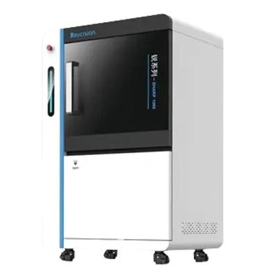
SHARP 1000 Multimodality Image Guided Precision Radiation System
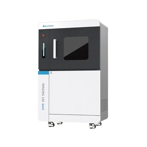
IMAGING 100 In Vivo/Ex Vivo Micro-CT Imaging System
SHARP 200 CT Guided Radiation System
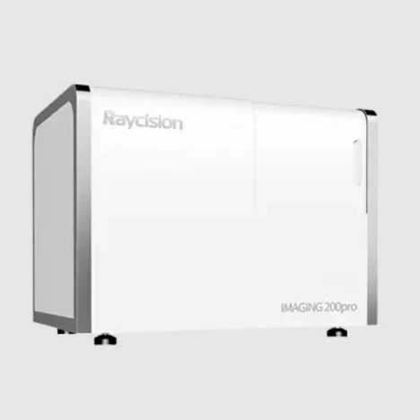
IMAGING 200 Pro 3D In Vivo Optical Molecular Imaging System
IMAGING 200 2D In Vivo Optical Molecular Imaging System
SHARP 100pro X-Ray Image Guided Radiation System
OTHER APPLICATIONS OF PRECLINICAL IMAGING AND RADIATION SYSTEMS
Cell and Molecular Biology
Material Science
Infectious Diseases
Raycision offers cutting-edge imaging technology and image guided radiation system for preclinical studies. Our radiation systems include everything from basic X-ray irradiators to multimodality image-guided precision radiation systems. The imaging systems offer a broad spectrum of capabilities extending from micro-CT imaging to optical molecular imaging. Whether for preclinical imaging or small animal radiation therapy, we are committed to providing products with exceptional performance and reliability.


Sign in to leave a comment.