Micro-CT and optical molecular imaging are powerful tools in cardiovascular research, each offering unique capabilities that enhance our understanding of radiation and cardiovascular diseases, development, and treatment. Here are some key applications of these imaging modalities in cardiovascular research:
Micro-CT Imaging
Vascular Morphology and Anatomy
3D Visualization: Micro-CT provides high-resolution, three-dimensional images of the vascular system, allowing detailed visualization of blood vessels, including small capillaries and complex vascular networks.
Quantitative Analysis: It enables precise measurement of vessel diameter, wall thickness, and branching patterns, which are crucial for studying vascular remodeling and angiogenesis.
Cardiac Structure and Function
Heart Morphology: Micro-CT imaging can provide detailed images of the heart’s anatomy, including the chambers, valves, and myocardium, aiding in the study of congenital heart defects and cardiomyopathies.
Functional Assessment: When combined with contrast agents, micro-CT can be used to assess cardiac function, including ventricular volumes and ejection fraction.
Vascular Development and Angiogenesis
Developmental Studies: Micro-CT is used to study the development of the vascular system in embryonic and postnatal stages, providing insights into normal and abnormal vascular development.
Angiogenesis Research: It allows for the evaluation of new blood vessel formation in response to therapies or in disease models.
Optical Molecular Imaging
Molecular and Cellular Processes
Gene Expression: Optical molecular imaging techniques such as bioluminescence and fluorescence imaging can visualize and quantify gene expression in the cardiovascular system, providing insights into molecular pathways involved in disease.
Cell Tracking: It allows for the tracking of labeled cells, such as stem cells or immune cells, to study their migration, homing, and integration in cardiovascular tissues.
Inflammation and Immune Response
Inflammatory Markers: Optical imaging can be used to detect and quantify inflammatory markers in the cardiovascular system, aiding in the study of inflammation’s role in diseases like atherosclerosis and myocarditis.
Immune Cell Dynamics: It enables the visualization of immune cell behavior in real-time, providing insights into their role in disease progression and response to therapies.
RAYCISION IMAGING AND RADIATION EQUIPMENTS
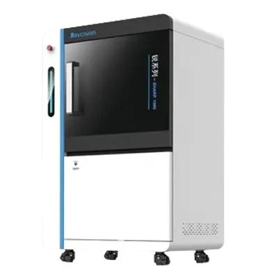
SHARP 100pro X-Ray Image Guided Radiation System
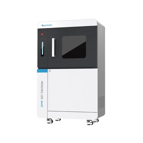
IMAGING 100 In Vivo/Ex Vivo Micro-CT Imaging System
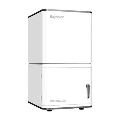
IMAGING 200 Pro 3D In Vivo Optical Molecular Imaging System
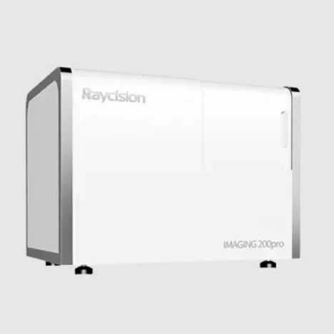
IMAGING 200 2D In Vivo Optical Molecular Imaging System
SHARP 1000 Multimodality Image Guided Precision Radiation System
IMAGING 1000 Multimodality Precision Imaging System
OTHER APPLICATIONS OF PRECLINICAL IMAGING AND RADIATION SYSTEMS
Animal Models
Drug Development
Material Science
Radiation induced coronary artery disease is a concern in bone disease treatment due to unintended radiation exposure of coronary arteries, leading to atherosclerosis. Mitigation strategies include precise radiation targeting with advanced imaging systems and research into therapeutic prevention.
If you want to know more about cardiovascular x ray and heart disease chest x ray image guided radiation system, please visit our website.
Raycision offers cutting-edge imaging technology and image guided radiation system for preclinical studies. Our radiation systems include everything from basic X-ray irradiators to multimodality image-guided precision radiation systems. The small animal imaging systems offer a broad spectrum of capabilities extending from micro-CT imaging to optical molecular imaging. Whether for preclinical imaging or animal radiation therapy, we are committed to providing products with exceptional performance and reliability.


Sign in to leave a comment.