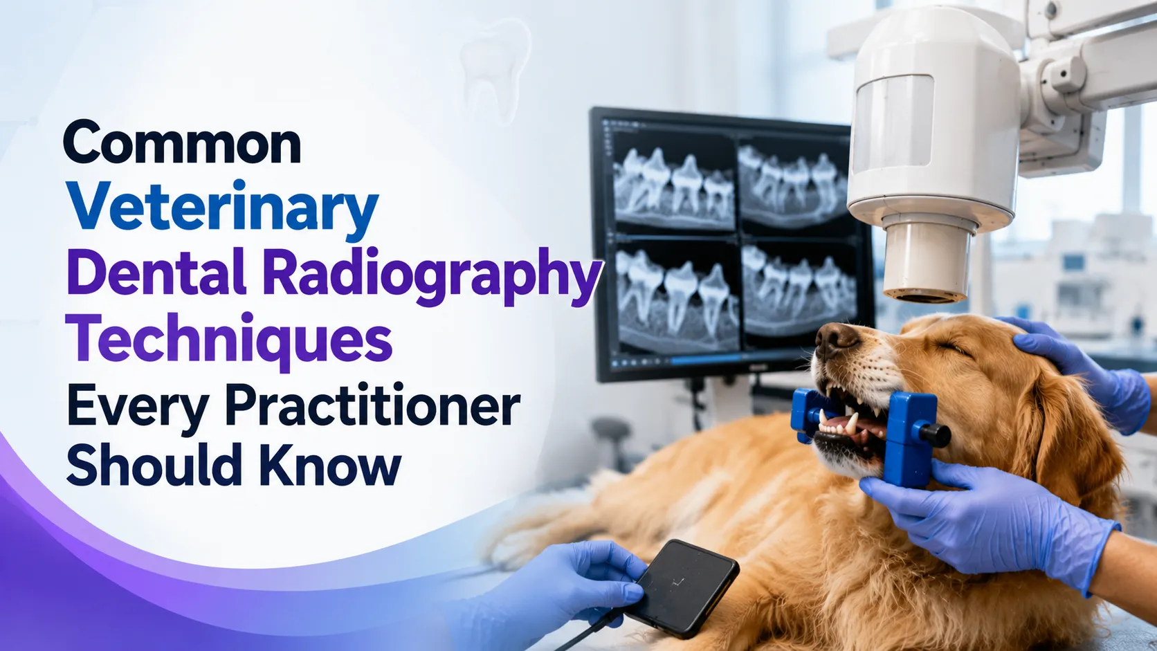

Dental disease is one of the most frequently diagnosed conditions in companion animals, yet much of it remains hidden beneath the gumline. A tooth can appear healthy during an oral examination while harboring root fractures, periodontal bone loss, or resorptive lesions that only dental radiographs can reveal.
That is why mastering veterinary dental radiography techniques is no longer optional for modern veterinary teams. It is an essential clinical skill that directly improves diagnosis, treatment planning, and patient outcomes.
For veterinarians and veterinary technicians, producing high-quality dental radiographs requires more than operating an X-ray unit. Correct positioning, understanding anatomical landmarks, selecting the appropriate imaging technique, and interpreting images accurately all contribute to successful patient care.
Whether you are building confidence with intraoral imaging or refining advanced positioning methods, improving your radiographic skills can significantly enhance the quality of your dentistry services.
At Vet and Tech, continuing education focuses on practical skills that veterinary professionals can immediately apply in clinical practice. Specialized webinars and expert-led training help practitioners strengthen their diagnostic abilities while keeping pace with advances in veterinary dentistry.
Why Are Veterinary Dental Radiographs Essential?
Dental radiographs provide information that cannot be obtained through visual examination alone. According to the American Veterinary Dental College (AVDC), most pathology associated with teeth occurs below the gumline, making dental imaging a standard component of complete oral examinations.
Common conditions identified through dental radiography include:
- Periodontal bone loss
- Tooth root abscesses
- Tooth resorption
- Retained tooth roots
- Jaw fractures
- Endodontic disease
- Developmental abnormalities
- Oral tumors involving bone
Research published in the Journal of Veterinary Dentistry has shown that full-mouth dental radiographs frequently identify clinically significant disease that would otherwise remain undetected during visual examination alone. This evidence reinforces why comprehensive dental imaging is considered the standard of care in companion animal dentistry.
Which Veterinary Dental Radiography Techniques Should Every Practitioner Know?
Several positioning techniques are used depending on the patient's anatomy and the tooth being evaluated. Choosing the correct approach minimizes distortion while producing diagnostic-quality images.
1. Parallel Technique
The parallel technique is considered the simplest positioning method and is primarily used for imaging the mandibular premolars and molars.
In this approach:
- The image receptor is positioned parallel to the tooth.
- The X-ray beam is directed perpendicular to both the receptor and the tooth.
- Minimal image distortion occurs.
Because the anatomy of the mandibular cheek teeth allows proper receptor placement, this technique consistently produces highly accurate images with excellent diagnostic value.
Best suited for:
- Mandibular molars
- Mandibular premolars
Advantages
- Excellent dimensional accuracy
- Easy positioning
- Minimal distortion
- High diagnostic reliability
2. Bisecting Angle Technique
The bisecting angle technique is the most widely used method in veterinary dental radiography because many teeth cannot accommodate the image receptor parallel to the tooth.
Instead, practitioners imagine an invisible bisecting line between the long axis of the tooth and the image receptor. The X-ray beam is directed perpendicular to this imaginary line.
This method is commonly used for:
- Maxillary incisors
- Maxillary canine teeth
- Maxillary premolars
- Maxillary molars
- Mandibular incisors
- Mandibular canine teeth
Although highly versatile, this technique requires precise positioning. Small changes in beam angulation can create elongated, shortened, or overlapping images that reduce diagnostic quality.
3. Extraoral Dental Radiography
While intraoral imaging remains the preferred standard, extraoral radiography has specific applications.
Practitioners may use it when:
- Patients have severe trauma
- Oral opening is limited
- Large breed anatomy prevents proper sensor placement
- Evaluating mandibular fractures
- Assessing temporomandibular joint disease
Extraoral imaging complements intraoral techniques rather than replacing them.
Comparison of Common Veterinary Dental Radiography Techniques
| Technique | Primary Use | Advantages | Limitations |
| Parallel Technique | Mandibular premolars and molars | Minimal distortion, highly accurate | Limited to specific anatomical regions |
| Bisecting Angle Technique | Most maxillary teeth and anterior mandibular teeth | Versatile, widely applicable | Requires precise positioning skills |
| Extraoral Radiography | Trauma, jaw assessment, limited mouth opening | Useful for complex cases | Lower detail for individual teeth |
What Makes a Dental Radiograph Diagnostic?
Obtaining an image is only the first step. Producing a diagnostic-quality radiograph depends on several technical factors working together.
Experienced veterinary dental teams consistently evaluate:
Correct Patient Positioning
Even slight patient movement can reduce image clarity. General anesthesia remains the recommended standard because it allows accurate positioning while minimizing patient stress.
Accurate Sensor Placement
Improper placement often results in:
- Cut-off tooth roots
- Cropped crowns
- Missing anatomical landmarks
- Image distortion
Sensor positioning should always prioritize visualization of the complete tooth, including surrounding alveolar bone.
Proper Beam Alignment
Incorrect beam angulation produces common imaging errors such as:
- Elongation
- Foreshortening
- Cone cuts
- Overlapping roots
Developing consistency with beam alignment takes practice but significantly improves diagnostic accuracy.
Common Positioning Mistakes and How to Avoid Them
Many image quality issues stem from simple positioning errors rather than equipment problems.
| Common Error | Likely Cause | Solution |
| Elongated tooth | Beam angle too shallow | Increase beam angulation |
| Foreshortened tooth | Beam angle too steep | Reduce beam angulation |
| Cone cut | Incorrect tube head alignment | Center the beam over the receptor |
| Blurred image | Patient movement | Improve stabilization under anesthesia |
| Overlapping roots | Incorrect horizontal angulation | Reposition beam before exposure |
Recognizing these patterns allows veterinary teams to make quick corrections without repeating multiple exposures.
Why Positioning Skills Matter More Than Equipment
Many practices invest in digital dental radiography systems expecting immediate improvements in image quality. While modern sensors and software certainly enhance workflow, technology cannot compensate for poor positioning.
Dr. Brett Beckman, Diplomate of the American Veterinary Dental College (AVDC), has frequently emphasized that accurate positioning is fundamental to obtaining diagnostic dental radiographs. Even advanced imaging systems rely on proper technique to produce clinically useful images.
For this reason, continuing education in positioning remains one of the highest-value investments for veterinary professionals involved in dentistry.
Veterinary technicians, in particular, play a central role in producing diagnostic images efficiently. As their positioning skills improve, practices often experience shorter anesthesia times, fewer retakes, and more confident treatment planning.
Professionals looking to strengthen these practical skills can benefit from Dental Radiography: Positioning with Perrone, an educational veterinary dentistry webinar available through Vet and Tech. The session focuses on real-world positioning techniques, common challenges, and practical methods that help veterinary teams capture consistent, high-quality dental radiographs in everyday practice.
How Can Practitioners Improve Their Dental Radiograph Interpretation Skills?
Capturing a clear image is only half the process. Interpreting what the image reveals is what guides clinical decisions. Every dental radiograph should be evaluated systematically rather than focusing only on the tooth that prompted imaging.
A consistent review includes:
- Crown integrity
- Root morphology
- Pulp chamber size
- Periodontal ligament space
- Lamina dura continuity
- Alveolar bone height and density
- Furcation involvement in multi-rooted teeth
- Periapical changes
- Adjacent teeth and supporting structures
This structured approach helps practitioners identify incidental findings that might otherwise go unnoticed.
According to Dr. Brook A. Niemiec, DVM, DAVDC, DEVDC, FAVD, one of the most respected authorities in veterinary dentistry, dental radiographs should be interpreted as carefully as thoracic or abdominal radiographs because clinically important pathology is often present without obvious oral signs. His work has consistently demonstrated that comprehensive radiographic assessment leads to earlier diagnosis and more appropriate treatment planning.
What Are the Most Common Dental Conditions Identified on Veterinary Radiographs?
Dental radiography plays a critical role in diagnosing conditions that are impossible to evaluate through visual examination alone.
Periodontal Disease
Periodontal disease affects the supporting tissues of the teeth and is considered the most common dental disease in dogs and cats. Radiographs reveal:
- Horizontal bone loss
- Vertical bone defects
- Furcation exposure
- Tooth mobility associated with bone destruction
Early detection allows clinicians to intervene before irreversible damage occurs.
Tooth Resorption
Tooth resorption is especially common in cats and may also occur in dogs.
Radiographs help determine:
- Type of lesion
- Severity
- Root replacement
- Appropriate extraction technique
Without dental imaging, these lesions may be mistaken for simple gingivitis or fractured teeth.
Endodontic Disease
A fractured tooth may appear uncomplicated externally while harboring pulp necrosis beneath the surface.
Radiographic findings include:
- Widened periodontal ligament space
- Periapical lucency
- Root resorption
- Open pulp chambers
These findings help determine whether extraction or endodontic treatment is appropriate.
Retained Tooth Roots
Retained roots are a frequent source of chronic pain and infection.
Dental radiographs confirm:
- Root fragments remaining after extraction
- Root ankylosis
- Root curvature
- Healing following surgical extraction
How Does Continuing Education Improve Veterinary Dental Imaging?
Veterinary dentistry continues to evolve with new imaging technologies, updated treatment protocols, and refined positioning techniques. Ongoing education helps veterinary professionals stay current while improving confidence in everyday practice.
Continuing education offers several practical benefits:
- Better diagnostic image quality
- Fewer radiograph retakes
- Reduced anesthesia time
- Increased confidence in interpreting findings
- More efficient workflow within the dental suite
- Improved communication between veterinarians and technicians
This is where dedicated training becomes especially valuable.
At Vet and Tech, veterinary professionals have access to educational resources developed specifically for clinical practice. Rather than focusing solely on theory, these programs emphasize practical techniques that can be implemented immediately in hospitals and specialty practices.
Frequently Asked Questions
1. Do all dogs and cats need dental radiographs during professional dental procedures?
Yes. The American Veterinary Dental College (AVDC) recommends full-mouth dental radiographs as part of a complete oral examination because a significant percentage of dental disease exists below the gumline and cannot be detected visually.
2. Which dental radiography technique is used most often?
The bisecting angle technique is the most commonly used method because it accommodates the anatomical challenges presented by the maxilla and anterior mandible. The parallel technique is primarily reserved for mandibular premolars and molars.
3. Can poor positioning affect diagnosis?
Absolutely. Incorrect positioning can create image distortion, hide pathology, or make healthy structures appear abnormal. Proper positioning reduces repeat exposures and increases diagnostic confidence.
4. Why is digital dental radiography preferred?
Digital systems provide:
- Faster image acquisition
- Improved image enhancement
- Lower radiation exposure compared with some traditional systems
- Easier image storage
- Better communication with clients
However, image quality still depends on proper positioning and technique.
5. How often should veterinary professionals receive dental radiography training?
Continuing education should be an ongoing process. As equipment, imaging software, and dental procedures evolve, regular training helps practitioners maintain consistent image quality and stay aligned with current standards of care.
Elevating Patient Care Starts with Better Imaging
Veterinary dental radiography has become one of the most valuable diagnostic tools in companion animal practice. From identifying hidden periodontal disease to detecting tooth resorption, fractures, retained roots, and endodontic pathology, high-quality dental radiographs support earlier diagnosis and more informed treatment decisions.
Yet even the best imaging equipment cannot replace strong technical skills. Consistent positioning, thoughtful image interpretation, and a commitment to continuing education are what separate adequate radiographs from truly diagnostic ones.
As Dr. Jan Bellows, DVM, DAVDC, DABVP, has often noted in his educational work, successful veterinary dentistry is built on accurate diagnosis before treatment begins. Dental radiography provides that diagnostic foundation.
Veterinary professionals who continue refining their imaging skills not only improve efficiency in the dental suite but also deliver a higher standard of care for every patient they treat. Through expert-led education, practical webinars, and career-focused learning opportunities, Vet and Tech supports veterinarians and veterinary technicians who want to advance their clinical expertise.
Resources like Dental Radiography: Positioning with Perrone help transform everyday imaging challenges into confident, consistent clinical practice, ultimately benefiting both veterinary teams and the animals they serve.


















Sign in to leave a comment.