Intertrochanteric fractures are a common type of hip fracture that occur in the upper part of the femur, specifically between the greater and lesser trochanter. These injuries are frequently seen in older adults, particularly those with osteoporosis, but they can also result from high energy trauma in younger individuals. Because the hip region plays a central role in weight bearing and mobility, effective management of these fractures is essential for restoring independence and preventing complications.
Understanding Intertrochanteric Fractures
Trochanteric Femur Nail is widely used in the surgical management of intertrochanteric fractures because it provides stable internal fixation aligned with the natural mechanical axis of the femur. Intertrochanteric fractures occur in the extracapsular region of the proximal femur and are often caused by a direct fall onto the hip. In elderly individuals with reduced bone density, even low energy trauma can result in significant displacement.
These fractures are typically classified as stable or unstable depending on the fracture pattern and the integrity of the medial and posterior cortical support. Stable fractures maintain structural continuity after reduction, while unstable fractures may involve comminution or loss of supportive bone structures. This classification plays a crucial role in determining the most appropriate surgical approach.
Without timely intervention, intertrochanteric fractures can lead to prolonged immobility. Extended bed rest increases the risk of complications such as deep vein thrombosis, pulmonary infections, muscle wasting, and pressure related skin injuries. Early surgical stabilization allows patients to mobilize sooner, significantly reducing these risks.
Biomechanical Principles of Intramedullary Fixation
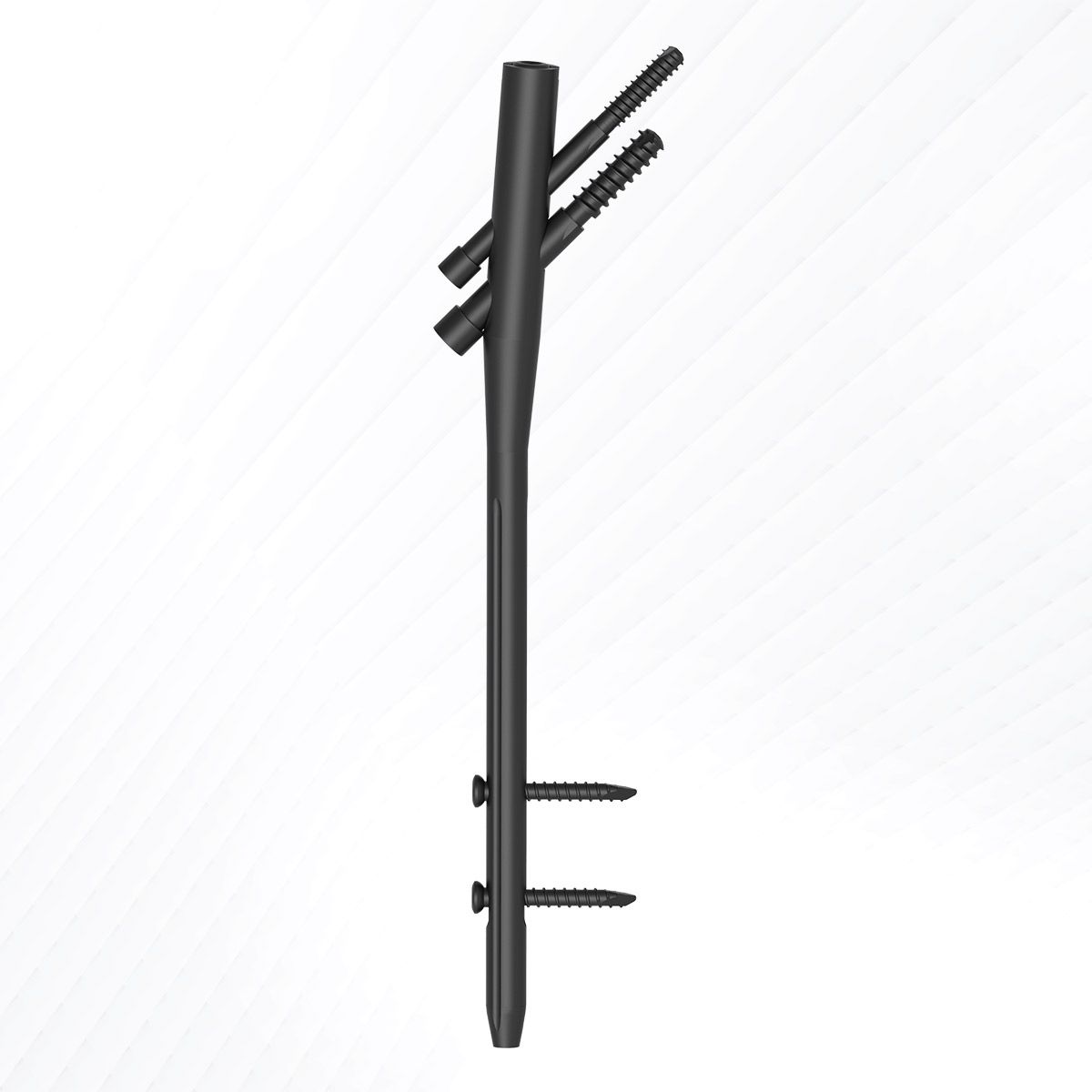
Intramedullary fixation involves inserting a metal implant into the medullary canal of the femur. Because the implant lies within the central axis of the bone, it provides effective load sharing during weight bearing. This alignment reduces the bending forces acting on the implant and supports more natural stress distribution across the fracture site.
The proximal component of the system typically includes a lag screw or blade that is inserted into the femoral head. This element secures the proximal fragment and allows controlled compression at the fracture site. Controlled compression can enhance stability and encourage bone healing by maintaining close contact between fracture fragments.
Distal locking screws prevent rotational instability and maintain overall alignment. Together, these components create a stable construct capable of withstanding the mechanical demands of early mobilization.
Advantages in Unstable Fracture Patterns
Unstable intertrochanteric fractures present unique challenges due to comminution and loss of medial support. Intramedullary systems are particularly beneficial in these cases because their central placement provides a shorter lever arm, which reduces stress on the implant.
In osteoporotic bone, achieving secure fixation can be difficult. The design of the proximal fixation element aims to improve purchase within the femoral head, decreasing the likelihood of mechanical failure. By maintaining alignment and resisting varus collapse, intramedullary fixation supports predictable healing even in complex fracture configurations.
Early weight bearing is often possible with stable internal fixation, depending on the surgeon’s assessment and the patient’s overall condition. This is especially important in elderly patients, where prolonged immobility can lead to rapid functional decline.
Minimally Invasive Surgical Technique
One of the notable benefits of intramedullary fixation is the relatively minimally invasive surgical approach. The implant is inserted through a small incision near the tip of the greater trochanter. In many cases, the fracture can be reduced without extensive exposure, preserving surrounding soft tissues.
Preservation of the soft tissue envelope is critical for maintaining blood supply to the fracture fragments. Adequate blood flow supports bone healing and reduces the risk of delayed union. Additionally, smaller incisions may result in reduced postoperative discomfort and quicker recovery in the early postoperative period.
Imaging guidance during the procedure helps ensure accurate placement of the implant and screws. Proper positioning is essential for maintaining alignment and preventing complications such as implant migration or joint penetration.
Rehabilitation and Recovery
Surgical fixation is only one part of successful fracture management. Rehabilitation plays a vital role in restoring function and independence. Following surgery, patients are typically encouraged to begin gentle hip and knee range of motion exercises under professional supervision.
Weight bearing is progressed gradually, based on fracture stability and individual healing response. Early mobilization supports muscle strength, joint flexibility, and overall cardiovascular health. It also contributes to improved mental well being by promoting independence and reducing the sense of prolonged illness.
Long term outcomes depend on several factors, including fracture severity, surgical accuracy, bone quality, and adherence to rehabilitation protocols. With appropriate management, many patients regain satisfactory mobility and return to daily activities.
Clinical Decision Making
Although intramedullary fixation is widely used, treatment must always be individualized. Surgeons evaluate patient age, bone density, fracture classification, and medical comorbidities before deciding on the most appropriate fixation method.
Preoperative planning and precise intraoperative technique are critical for minimizing complications. Postoperative follow up allows monitoring of fracture healing and early identification of any concerns.
In certain clinical scenarios, alternative fixation methods may be considered. Therefore, comprehensive assessment and careful surgical judgment remain central to effective fracture management.
Conclusion
Intertrochanteric fractures require timely stabilization to restore mobility and reduce the risks associated with prolonged immobilization. Intramedullary fixation offers biomechanical strength, central load sharing, and a minimally invasive approach that supports early rehabilitation.
Understanding the principles behind this surgical method helps patients and caregivers appreciate the rationale for its use. With proper planning, skilled surgical execution, and structured rehabilitation, many individuals can achieve meaningful recovery and regain functional independence after an intertrochanteric fracture.
Sign in to leave a comment.