For over a century, the fundamental design of the optical microscope remained largely unchanged. It was an instrument defined by solitary use, where a single observer would hunch over eyepieces to interpret the secrets of cellular biology. However, the rapid digitalization of healthcare has forced a re-evaluation of these traditional tools. As laboratories face increasing pressure to deliver faster results and facilitate remote consultations, the industry is witnessing a pivot toward smart, integrated devices. Leading this charge is the tablet microscope, a sophisticated fusion of high-end optics and digital computing that is reshaping the way pathology and biology are practiced today.
Overcoming the Limitations of Analog Systems
The traditional optical workflow, while reliable, is fraught with inefficiencies that modern labs can no longer afford. The physical act of microscopy has historically been a major source of occupational strain. Pathologists and technicians frequently report neck and back pain from hours spent in rigid positions, a factor that can contribute to fatigue and potential errors in diagnosis. Furthermore, the analog nature of these older devices creates a silo effect; if a difficult case requires a second opinion, the physical slide often has to be transported to another location, introducing delays that can critically impact patient care.
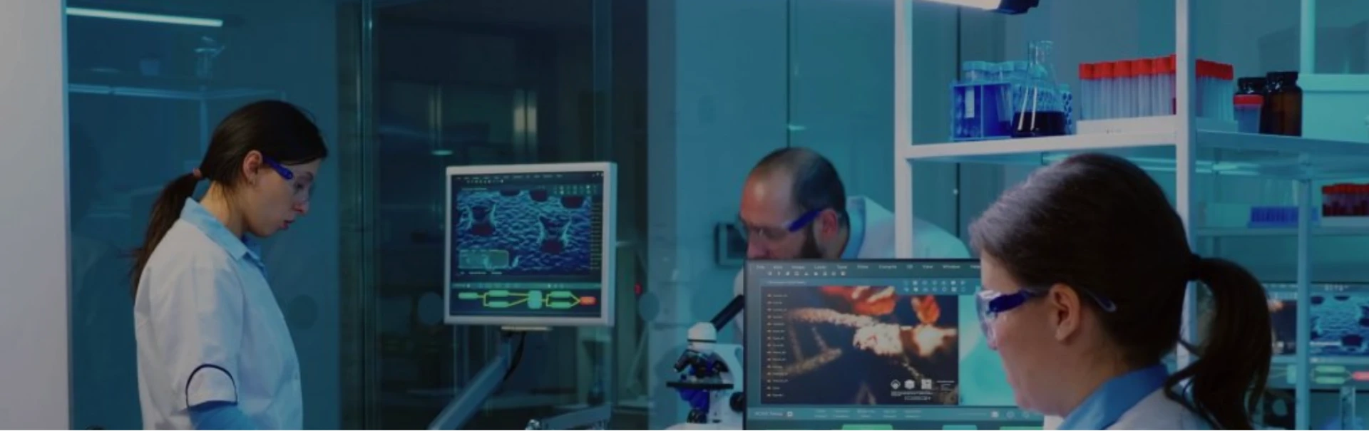
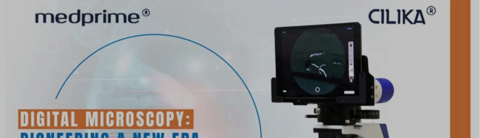
Digital integration solves these ergonomic and logistical hurdles simultaneously. By replacing or augmenting the standard viewing head with a high-definition touchscreen interface, these new devices allow users to view specimens in a "heads-up" manner. This shift not only improves posture and comfort but also drastically expands the field of view. The ability to manipulate the image directly on a screen—zooming, panning, and adjusting contrast with touch gestures—brings a level of interactivity that analog systems simply cannot match. It transforms the microscope from a passive viewing tool into an active data generation hub.
The Role of Connectivity in Modern Diagnostics
The true power of this technology lies in its ability to connect. In an era where telepathology is becoming essential, particularly in underserved or remote regions, the ability to transmit live images is revolutionary. A tablet microscope functions as a standalone communication unit, allowing laboratory technicians in peripheral centers to connect instantly with specialists in major hospitals. Through live streaming or high-resolution image capture, a pathologist hundreds of miles away can view the sample as if they were sitting in the lab itself, guiding the technician to specific areas of the slide and providing an immediate diagnosis.
This connectivity also streamlines the workflow within the lab itself. In the past, archiving a sample meant storing fragile glass slides in massive physical cabinets, where they were prone to breakage or degradation over time. Digital microscopy allows for the creation of virtual slide libraries. Images and videos can be saved directly to a server or cloud storage, attached to patient electronic health records, and retrieved instantly for future comparison. This digital trail is invaluable for tracking disease progression and ensures that the patient's data is comprehensive and easily accessible to their entire care team.
Enhancing Education and Training Efficiency
Academic institutions have been among the fastest adopters of this technology, recognizing its potential to democratize learning. In a conventional biology lab, an instructor must move from student to student, peering into individual eyepieces to verify what is being seen. It is a slow, repetitive process that often leaves students unsure if they have identified the correct structure.
With screen-based systems, this dynamic is inverted. An instructor can project a live feed to a larger display or view the student’s screen directly, ensuring that the entire group is looking at the same anomaly simultaneously. This collaborative environment fosters better engagement and faster learning curves. Students can capture images of their findings, annotate them directly on the screen, and submit them for review, creating a seamless loop of feedback and assessment that prepares them for a digital-first professional environment.
The Future of Smart Imaging and AI Integration
As we look toward the next decade of medical diagnostics, the hardware we use today will serve as the foundation for artificial intelligence applications. The move toward screen-based data collection is the necessary first step in enabling AI-assisted pathology. A digital feed allows software algorithms to run in real-time, potentially assisting pathologists by pre-screening samples for specific cell types, counting blood cells automatically, or flagging suspicious areas for closer review.
The transition to these intelligent systems is being driven by forward-thinking manufacturers who understand that the future of the lab is digital. Companies are now focusing on creating devices that are not just optical tools, but comprehensive diagnostic platforms. This evolution ensures that even smaller laboratories can access world-class diagnostic capabilities without the need for massive infrastructure investments. By prioritizing user-friendly interfaces and robust connectivity, these innovations are bridging the gap between high-tech research and everyday clinical practice.
Pioneering Reliable Solutions for Global Labs
The market for these advanced diagnostic tools is competitive, but certain innovators stand out by focusing on the specific needs of the pathology community. The demand is for equipment that is rugged enough for field use yet sensitive enough for critical diagnosis. Meeting this demand requires a deep understanding of both optical physics and software engineering. This is the precise intersection where industry leaders are making their mark, developing products that simplify complex workflows while maintaining the highest standards of image fidelity.
For laboratories looking to future-proof their operations, the choice of equipment partner is critical. It is essential to select devices that offer seamless integration, ongoing support, and the reliability required for clinical environments. As the adoption of digital pathology becomes the standard rather than the exception, the solutions provided by Medprime Technologies continue to set the benchmark for quality and innovation in the field.


Sign in to leave a comment.