Artificial intelligence in ophthalmology brings a reassuring shift with the use of rich digital imaging data, including fundus photographs, optical coherence tomography (OCT), and visual field tests. This data supports disease diagnosis, patient care, and treatment planning. AI can recognize patients with preventable vision loss earlier. It also leads to the optimized allocation of medical resources through precise predictions and personalized interventions. AI applications in ophthalmology encompass medical pathology, imaging, analyzing large-scale patient data, and telemedicine services.
Let’s explore in detail how artificial intelligence holds immense potential to transform ophthalmic care by providing clinicians with data-driven, objective assessments. The blog outlines advancements and challenges of applying AI to major domains in ophthalmology.
The Progress so far
Significant progress has already been made. In 2018, IDx-DR became the first FDA-approved autonomous AI system for detecting diabetic retinopathy, marking a significant milestone. Since as early as 2008, AI has been applied across ophthalmology for screening and diagnosis of conditions such as diabetic retinopathy, glaucoma, retinopathy of prematurity, and keratoconus, as well as for treatment planning, outcome prediction, and workflow optimization. In January 2025, AI Optics received FDA 510(k) clearance for its Sentinel Camera, a handheld retinal imaging device built for improving retinal screening at the point of care. The portable device captures high-quality images without eye dilation and aims to support early detection of retinal diseases.
Yet, the path to responsible AI adoption in ophthalmology does not come without challenges. Voluminous high-quality training data are crucial to a model's performance. It is still difficult to uphold clinical trust and patient safety while simultaneously addressing ethical obligations, technological constraints, and regulatory complexity. Bias in AI systems raises questions about transparency and accountability. Due to AI inaccuracies, often referred to as hallucinations, there is a need for human-in-the-loop supervision to provide meticulous validation and oversight in clinical decision-making.
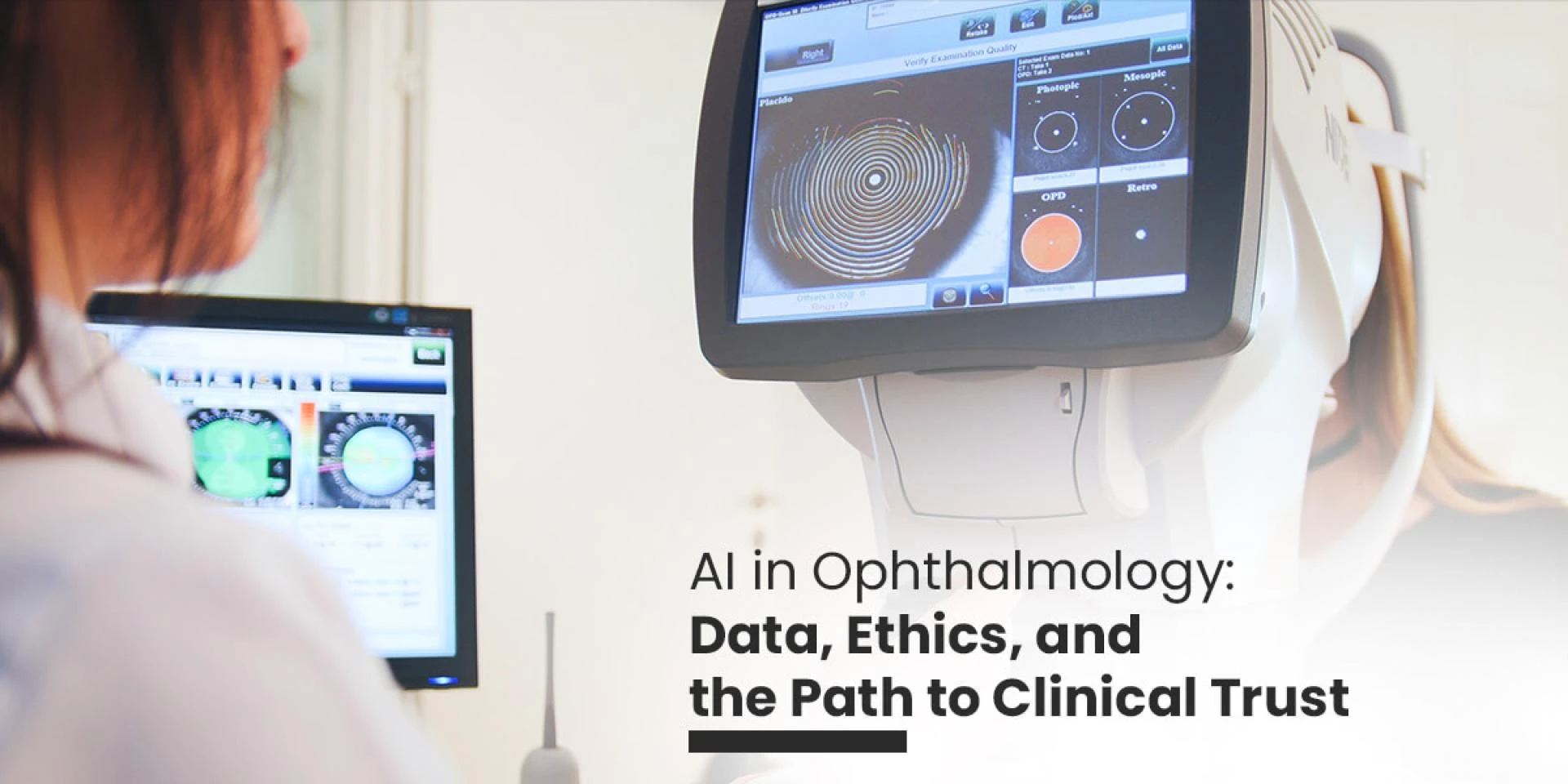
Imaging Enhancement and Analysis
Deep learning (DL) has brought advancements in ophthalmic imaging analysis. In terms of OCT, AI algorithms are available to improve image quality, extract clinically meaningful biomarkers, and perform semantic segmentations. A group of researchers shared that DL can effectively minimize OCT image noise and artifacts. Furthermore, Noise2Noise algorithms denoise images by leveraging the intrinsic redundancy in repeated scans.
AI has also made it feasible to automate segmentation of retinal layers and quantification of morphological features such as drusen and fluid. Researchers also agreed that AI-powered segmentation of choroidal neovascularization lesions in OCT angiography closely matches clinician-led assessments. The application of deep learning (DL) to visual field testing, ultrasonography, and other ophthalmic imaging modalities presents strong potential for extracting clinically meaningful biological markers.
Apart from screening, AI also aids treatment decisions by predicting disease progression. However, possible constraints, such as the diversity & quality of training data, as well as the lack of stringent clinical validations, become a challenge. Under the guidance of board-certified ophthalmologists, retina and glaucoma specialists, optometrists, and trained ocular imaging professionals, AI systems can address such limitations and set new standards for precise, evidence-based ophthalmic care.
Challenges and Considerations
There is no denying that AI holds promise for strengthening ophthalmology and optometry; however, its challenges must be addressed. The fundamental limitation is related to the representativeness and quality of the datasets used to train AI algorithms. There is a need for high-quality, diverse, and large datasets.
Diabetic retinopathy (DR)
Diabetic retinopathy (DR) is a leading cause of vision loss and preventable blindness worldwide. It affects approximately one-third of individuals with diabetes. Early detection and timely intervention can help prevent irreversible visual impairment. However, manual screening of retinal images for DR is labor-intensive, time-consuming, and costly. Thus, it requires specialized clinical expertise. These limitations have hindered the widespread implementation of large-scale DR screening initiatives. The model's performance can differ depending on the camera used and image quality.
Glaucoma
Glaucoma is one of the leading causes of irreversible blindness globally. Early diagnosis remains decisive to initiate treatment and prevent loss of sight. Nevertheless, glaucoma screening remains challenging as early-stage disease is often asymptomatic and diagnosis needs different specialized tests performed by trained clinicians. Fundus photography and the use of AI also face specific challenges for glaucoma screening.
The appearance of the optic disc varies due to factors such as image quality, acquisition devices, and refractive errors. Such factors can limit AI performance in anatomically complex or low-quality cases like tilted discs.
AI models can deliver real-world screening results by combining fundus images, OCT, and visual field data. With prospective validation and training data, fundus-based AI has strong potential for scalable, cost-effective early glaucoma detection.
This means that you need an expert training data service provider by your side to deliver comprehensive datasets that enable the development of algorithms capable of generalizing across diverse clinical scenarios. A data annotation company also helps establish standard regulatory guidelines and protocols to maintain safety and ethical implementation of DL applications in clinical settings.
Safeguarding Patient Privacy and Data Integrity
To handle sensitive data, including retinal images and personal health records, strict security measures are indispensable in ophthalmology. It aids in preventing misuse and unauthorized access. Stringent data protection laws, such as the GDPR and HIPAA, should be followed by healthcare researchers and practitioners. These frameworks need informed consent, secure data handling, access controls, and patient confidentiality protections.
As AI adoption in ophthalmology grows, safeguarding patient data remains a core ethical and regulatory responsibility. Trusted data annotation service providers implement privacy pipelines that enforce secure access, maintain audit trails, and ensure consent-aware dataset management. With the evolution of ophthalmology, collaborative efforts of healthcare professionals, scientists, patients, policymakers, and data annotation companies ensure that advancements in technology improve patient care without compromising patient trust or privacy in the medical community.
Bias and Fairness
It seems a prerequisite to create datasets that represent global diversity, as these reduce algorithmic bias. This ophthalmology advancement is supported by randomized controlled trials (RCTs) that assess AI systems under real clinical conditions, with increasing adherence to CONSORT-AI guidelines to maximize transparency, reproducibility, and clinical credibility.
Data annotation service providers help mitigate bias across the AI lifecycle. At the pre-processing stage, they support with equitable datasets to ensure consistent labeling standards, demographic balance, and representative pathology coverage. Post-processing reviews further evaluate model outputs across subpopulations, allowing the identification and correction of residual performance disparities.
In order to assist AI developers in developing systems that satisfy performance standards and foster fair access, interpretability, and trust in a variety of healthcare settings, data annotation partners provide a comprehensive package of accurate labels, expert-reviewed ground truth, and flexible annotation strategies.
Conclusions
Artificial intelligence supports ophthalmology to determine clear clinical value for glaucoma assessment, retinal screening, and multimodal imaging analysis. The real-world success of a model is not only based on advanced algorithms, as data diversity, integrity, and quality are equally vital. Therefore, it is imperative to deal with challenges like validation, bias, and patient privacy for safer AI adoption.
High-fidelity training data, human-in-the-loop validation, and expert-led annotation help turn AI systems into trusted clinical tools. Hence, a specialized data annotation partner will be critical to deliver responsible, compliant, and scalable AI solutions.


Sign in to leave a comment.