
Hearing about a breast lump can immediately raise questions, doubts, and quiet worries. Earlier, getting clear answers often took time, with multiple visits and unclear explanations along the way. Today, that experience has become far more structured and reassuring. Better imaging tools and intelligent systems now support clearer conversations between doctors and patients.
This blog explains how modern imaging and AI are changing the way breast lumps are evaluated, how decisions are made with greater confidence, and why these advances matter for everyday patients seeking clarity and peace of mind.
Why Breast Lump Diagnosis Feels Different Today
Breast lump diagnosis has become calmer and structured compared to the past. Doctors now rely on clearer information from the start, which helps avoid rushed conclusions and unnecessary worry. Early screening and awareness have also encouraged people to seek medical advice sooner.
Earlier breast lump treatment in Ahmedabad awareness campaigns and routine screening have encouraged people to come in sooner, often before symptoms escalate. Instead of assumption-based physical checks alone, image-backed insights now guide conversations. Accuracy at the first stage matters because it sets the tone for everything that follows. Technology has also shifted expectations, with patients wanting clarity, timelines, and understandable explanations from the start.
What Has Improved in Breast Imaging Over the Last Few Years
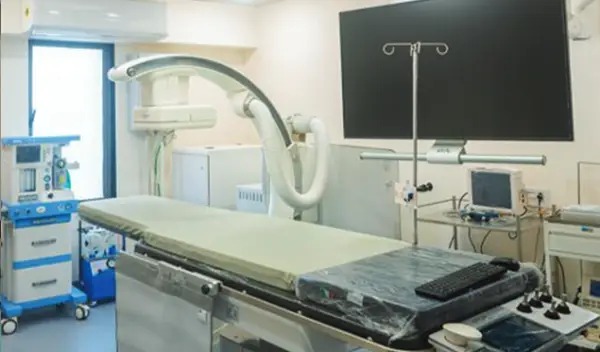
Breast imaging has quietly evolved, but the impact is significant. Modern machines capture finer details, helping doctors understand tissue patterns more clearly. This improvement reduces doubt and speeds up the overall process. Higher image resolution and faster reporting mean fewer follow-up scans are needed. Software-supported image analysis adds another layer of review, supporting clinical judgement.
Many imaging setups are also designed with comfort in mind, making the experience less intimidating. Together, these changes create smoother evaluations with less repetition and more confidence for both doctors and patients.
How AI Supports Doctors in Identifying Breast Lumps More Clearly
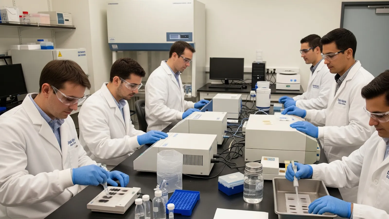
AI does not replace doctors; it supports them. Its role is to assist with pattern recognition and highlight areas that deserve closer attention. This support helps reduce variability and missed details.
AI-assisted tools compare imaging data against large reference patterns, helping spot subtle differences that may not stand out visually. This consistency supports clearer reporting across cases. By handling repetitive analytical tasks, AI allows doctors to focus more on clinical decision-making and patient communication, which improves overall care quality.
Types of Imaging Tests Commonly Used to Check Breast Lumps
Different imaging tests serve different purposes, and choosing the right one depends on what doctors need to understand. Each method offers unique information that contributes to a complete picture.
Ultrasound helps assess tissue characteristics and is often the first step. Mammography provides insight into structure and density, especially useful for screening. MRI is reserved for complex or unclear findings where deeper detail is needed. Image-guided procedures improve accuracy when sampling or targeted evaluation is required, reducing uncertainty during diagnosis.
How Imaging Results Help Decide the Right Treatment Approach
Imaging does more than identify a lump; it guides what happens next. Clear visuals help doctors understand size, location, and behaviour, which directly influences treatment decisions.
With precise imaging, doctors can differentiate between cases that need observation and those requiring intervention. This clarity supports personalised planning rather than one-size-fits-all decisions. In many diagnostic pathways, imaging insights naturally shape discussions around Breast lump treatment helping align medical recommendations with individual needs and comfort levels.
How Advanced Imaging Also Supports Other Women’s Health Treatments
The best uterine fibroids treatment in Ahmedabad now benefits from the same advanced imaging principles used in breast care. Detailed visual information allows doctors to understand fibroid size, location, and impact before deciding on the most suitable care pathway.
The benefits of advanced imaging extend beyond breast care. Similar principles are used across women’s health to support accurate diagnosis and thoughtful treatment planning. Imaging plays a key role in gynecological assessments, helping doctors map structures and plan interventions with greater confidence.

What Patients Can Expect During an Imaging-Based Breast Evaluation
An imaging-based evaluation is usually straightforward and well-explained. The process begins with a consultation to decide which scan is most suitable.
During imaging, patients often notice improved comfort and shorter scan times. Results are reviewed carefully and explained in clear language, with visual references when helpful. Based on findings, doctors outline the next steps, which may include monitoring, further tests, or discussions around Ahmedabad, depending on individual results.
What Patients Usually Want to Know About AI and Imaging
Most questions revolve around safety, reliability, and trust. Modern imaging techniques follow strict safety standards, and AI tools are designed to support, not override, medical judgment.
Data privacy is taken seriously, with ethical use frameworks guiding AI systems. These tools are tested across age groups to ensure consistent performance. Importantly, final interpretations always involve specialists, ensuring technology remains a support system rather than a decision-maker.
Frequently Asked Questions
1. Is AI-supported imaging safe for breast lump diagnosis?
AI-supported imaging follows strict safety protocols and works alongside standard imaging techniques. It does not expose patients to additional risk. Doctors use it as an assistive tool, ensuring that clinical judgement remains central to diagnosis and care decisions.
2. What is the most effective treatment for uterine fibroids?
The most effective fibroid treatment is chosen after a proper clinical assessment. The best uterine fibroids treatment in Ahmedabad aims to reduce symptoms while preserving the uterus.
3. How accurate are imaging results compared to older methods?
Modern imaging combined with AI offers more consistent and detailed insights than older techniques. Improved resolution and analysis reduce variation in interpretation. This leads to clearer conclusions and more confident treatment planning.
Conclusion
AI and advanced imaging have changed how breast lumps are evaluated, making the process clearer, calmer, and more reliable. From early detection to thoughtful treatment planning, these tools support informed decisions without removing the human element. Patients benefit from better explanations, fewer delays, and more confidence in what comes next.
When combined with experienced medical judgement, technology becomes a steady guide rather than a source of confusion. At Dev Hospital, this balanced approach helps patients feel supported, informed, and reassured throughout their diagnostic journey.


Sign in to leave a comment.