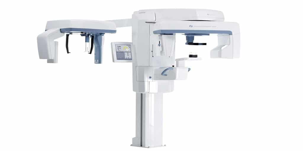
Dental CBCT (cone beam computed tomography) and 3D imaging are quickly becoming the new standard of care for dentists and dental specialists. Dentists and dental specialists can utilize CBCT technology in the production of 3D images and to create cross-sectional images, in order to improve diagnostic capabilities and service offerings
Dental cone beam imaging gives dental professionals the unprecedented ability to evaluate a patient’s dental health in three dimensions. Dental CBCT technology delivers high-quality imaging that allows for superior visualization of anatomical structures. It also provides powerful image management capabilities that allow for superior treatment planning. Here is everything a dental professional needs to know about dental CBCT technology.
Voxels, Field of View, and Image Acquisition
Two-dimensional digital images are made up of pixels which are small units of the area with height and width. The greater the number of pixels an image contains, the greater the resolution.
In three-dimensional imaging, pixels are replaced with voxels – small units with volume (length, height, and width) This adds a third dimension to the image. Voxels are usually represented as a cube or box with sides of 1-2mm in length. As voxel size decreases, spatial resolution increases, but so does the noise of the image.
Image quality is dependent on the field of view (FOV) scan time, the number of basis images, type of detector, tube voltage, and amperage. FOV widths can range from 3cm to as high as 24cm. Operators can choose a small, medium, or large FOV image. The appropriate FOV size is largely determined by the applications or procedures for which they will be used. Most medium or large FOV dental CBCT systems can collimate down to achieve smaller FOV sizes as necessary.
A CBCT scan captures anatomical data in one rotation, typically between 10 and 40 seconds; patients may either stand or sit, depending on the unit. The system’s software then reconstructs the data to show dimensionally accurate images of the scanned area in different planes. Creating the 3D images typically takes between 20 seconds and 6 minutes.
The software also allows for the manipulation of these images in order to show different slices at different angles, varying depths and thickness, 3D reconstructions, and some soft tissues without distortion.
Applications
CBCT images can assist in treatment planning for a variety of dental specialties, including oral and maxillofacial surgery, orthodontics, endodontics, and periodontics. Research also discusses the case-specific use of CBCT imaging for TMJ evaluation and airway analyses for obstructive sleep apnea.
In addition to providing a broader range of clinical applications, dental CBCT also helps enhance patient communication and treatment acceptance. When patients can visualize their anatomy in three dimensions, it helps them better understand their dental conditions and accept treatment recommendations.
Radiation Dosage and Exposure
CBCT requires an increased radiation dose compared to panoramic radiographs. Oral health professionals need to reduce dosages to a level at which diagnostic quality is not affected. They must also adhere to the ALARA (As Low As Reasonably Achievable) principle and exposure guidelines as set forth by the ADA and FDA. Most CBCT machines in the marketplace today design their equipment to specifically address ALARA, but offering individualized selection of exposures, equipment maintenance, and an ongoing quality assurance program.
Cost
Market demand has increased the affordability of dental CBCT systems, although these machines still typically represent a significant investment for most practices. A new CBCT system will typically range between $75,000 to $100,000, depending on the model and manufacturer. However, certified pre-owned systems also deliver a great deal of value to those looking for options that afford the latest technology and a high level of patient care.
There are many high-quality pre-owned systems available for purchase, often at up to 50% less than new prices. A pre-owned dental cone beam system gives the ability to provide patients with the benefits of new cone beam technology at significant cost savings.
Renew Digital, for example, is a leading provider of certified pre-owned dental imaging equipment. They offer a wide range of pre-owned dental CBCT machines at significantly discounted prices, often 30% to 50% off the price of new. They ensure high quality through a rigorous system of testing and inspection, delivering systems that will work safely and efficiently for years to come.
You can learn more online at RenewDigital.com or contact them directly at 888-246-5611.
For more information about Extraoral Bitewings and Sirona Panoramic Please visit: Renew Digital, LLC.