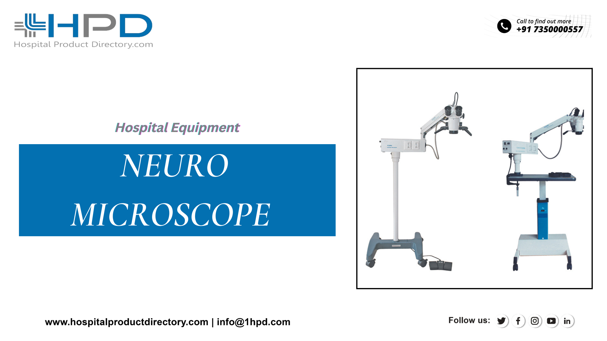
Deliberate your choices with your doctor when it comes to the kinds of tumor elimination surgeries using Neuro Microscopes supplied by Neuro Microscope Suppliers. Some choices may comprise:
Brainstem Surgery: Brainstem surgery is a kind of neurosurgery that includes operating on the brain stem, the low part of the foot of the brain that attaches the upper brain to the spinal cord. The brainstem influences many important purposes, including breathing, heart rate, and blood pressure, as well as the muscles used for eating and speaking. Brainstem surgery is naturally done to eliminate brain tumors, cavernous deformities, or other irregularities. This kind of surgery is very intricate and must be carried out with attention, as the brainstem is a subtle and critical part of the nervous system. It is typically only completed when other treatments are not conceivable or have been ineffective.
Cranioplasty: Cranioplasty is a clinical procedure in which a segment of the skull that has been detached or damaged is substituted or repaired. This procedure is often done after brain surgery to eliminate a brain tumor or to restore a skull fracture. It may also be completed to correct a malformation or to recover the appearance of the head. Cranioplasty can be done using a diversity of resources, including bone implants, synthetic materials, and metallic plates. The precise method used will be contingent on the location and size of the flaw and the patient's individual needs.
Craniotomy: To complete a craniotomy, your neurosurgeon will make an opening using a Neuro Microscope in the scalp to reach the tumor. If conceivable, your surgeon will perform a minimally invasive approach to cut healing and craniotomy retrieval time.
Awake Craniotomy: An awake craniotomy is a clinical procedure in which the patient is kept conscious and alert during a portion of the surgery. This is characteristically completed when the surgeon needs to access parts of the brain that control purposes such as speech, movement, or feeling, and it is essential to test these functions during the operation to safeguard that they are not damaged.
During an awake craniotomy, the patient is naturally given a local anesthetic to numb the part where the cut will be made, and the rest of the body is kept numb using general anesthesia. The patient is then woken and asked to perform various errands, such as talking, moving exact parts of the body, or recognizing objects, while the surgeon works on the brain. The patient is usually able to connect with the surgeon and the surgical team during the procedure, and they may be given medicines to help them relax and stay contented.
Awake craniotomy is a compound and highly dedicated surgical procedure and it is typically only done by experienced neurosurgeons. It is an important tool for diminishing the risk of problems and safeguarding the best possible outcome for the patient.
Endonasal Endoscopy: Endonasal surgery is a kind of surgical procedure that is done through the nostrils, without the need for external cuts. It is typically used to treat circumstances that affect the nasal hollow, sinuses, and skull pedestals, such as nasal growths, nasal tumors, and nasal fractures.
Endonasal surgery is typically done using dedicated instruments, such as endoscopes (long, reedy pipes with a light and a camera on the end), that are implanted through the nostrils and into the nasal hollow. The surgeon uses the endoscope to imagine the inside of the nose and sinuses and to complete the surgical procedure.
Endonasal surgery has several advantages likened to traditional open surgery, including a briefer retrieval time, less pain, and less scarring. It is also less aggressive, as it does not require any external cuts, and it allows the surgeon to contact the nasal cavity and sinuses more directly.
Image-guided Tumor Resection: Image-guided tumor resection is a surgical process in which the surgeon uses dedicated imaging techniques, such as magnetic resonance imaging (MRI) or computed tomography (CT) examinations and the use of a Neuro Microscope bought from Neuro Microscope Suppliers, to steer the elimination of a tumor or other mass. This permits the surgeon to envisage the site and degree of the tumor like a “GPS” chart of a patient’s brain and to recover the chance to eliminate the target more precisely and completely.
Guided tumor resection is an imperative tool for refining the result of tumor surgery and it is an important part of many cancer treatment plans. Patients need to work carefully with their healthcare team to mature a treatment plan that is suitable for their requirements.
Minimally Invasive Skull Base Surgery: Minimally invasive skull base surgery is a kind of surgical process that is done through small incisions, using dedicated instruments, Neuro Microscopes, and techniques. Minimally invasive surgery aims to curtail the size of the cut and the amount of tissue that is disturbed, which can lead to less discomfort, faster retrieval, and a lower danger of problems.
Minimally invasive skull base surgery is used to treat circumstances that disturb the skull pedestal, which is the portion at the bottom of the skull where it encounters the spine. These ailments may comprise tumors, aneurysms, and other irregularities of the brain and blood vessels. Some shared instances of minimally invasive skull base surgery include endoscopic surgical treatment, micro neurosurgery, transnasal surgery, and transoral surgical treatment. These actions may be done using small cuts and specialized instruments, such as endoscopes and neuro microscopes supplied by Neuro Microscope Suppliers.


