
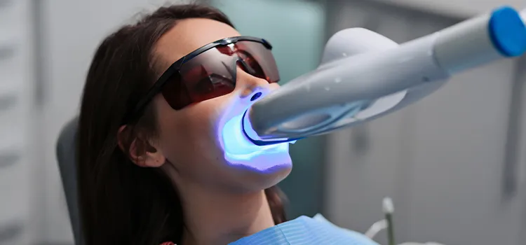
Dental X-rays done on equipment supplied by Dental X-Ray Suppliers support dentists envisage illnesses of the teeth and adjacent tissue that cannot be understood with a simple oral exam. They also aid the dentist to find and treat dental glitches early on, which can help save you money, needless uneasiness, and maybe even your life.
Kinds of Dental X-Rays
There are two chief kinds of dental X-rays: intraoral (entailing the X-ray film inside the mouth) and extraoral (requiring the X-ray film outside the mouth).
Intraoral X-rays are the most shared kind of dental X-ray taken. These X-rays deliver a lot of aspects and permit your dentist to find craters, check the fitness of the tooth root and bone adjacent to the tooth, check the position of emerging teeth, and monitor the overall fitness of your teeth and jawbone.
Extraoral X-rays display teeth, but their chief focus is the jaw and skull. These X-rays do not deliver the aspect found with intraoral X-rays and therefore are not used for noticing cavities or for recognizing glitches with individual teeth. Instead, extraoral X-rays are used to look for wedged teeth, screen development and expansion of the jaws about the teeth, and to recognize potential glitches between teeth and jaws.
Kinds of Intraoral X-Rays
There are numerous kinds of intraoral X-rays, each of which displays different facets of teeth.
Bite-wing X-rays display particulars of the upper and lower teeth in one part of the mouth. Each bitewing displays a tooth from its crown to around the level of the supportive bone. Bite-wing X-rays are used to notice deterioration between teeth and variations in bone density caused by gum illness. They are also useful in defining the correct fit of a crown (or cast refurbishment) and the marginal veracity of fillings.
Periapical X-rays display the entire tooth -- from the crown to outside the end of the root to where the tooth is fastened in the jaw. Each periapical X-ray displays this full tooth length and comprises all the teeth in one share of either the upper or lower jaw. Periapical X-rays are used to notice any irregularities in the root construction and nearby bone construction.
Occlusal X-rays are bigger and display full tooth growth and assignment. Each X-ray discloses the complete arch of teeth in either the upper or lower jaw.
Kinds of Extraoral X-Rays
Numerous kinds of extraoral X-rays are done on equipment bought from Dental X-Ray Dealers in India that your dentist may take.
Panoramic X-rays display the complete mouth area -- all the teeth in both the upper and lower jaws -- on a solitary X-ray. This kind of X-ray is valuable for noticing the location of fully arisen as well as developing teeth, can recognize impacted teeth, and aid in the analysis of tumors.
Tomograms display a specific layer or "slice" of the mouth while distorting out all other layers. This kind of X-ray is valuable for probing edifices that are problematic to see -- for example, because other edifices are in very close proximity to the structure to be watched.Cephalometric projections display the complete side of the head. This kind of X-ray is valuable for probing the teeth about the jaw and outline of the individual. Orthodontists use this kind of X-ray to mature their treatment plans.
0


Sign in to leave a comment.