Why better thermometry helps clinicians heat with purpose, not guesswork
It often comes down to a quiet moment in the treatment room.
Energy is being delivered. The treatment plan looks right. The target has been selected. But one question still decides whether the therapy stays helpful or becomes hazardous: Where is the heat actually going?
That is why temperature measurement in cancer thermal therapies matters so much. In hyperthermia and other heat-based cancer treatments, success is not just about raising temperature. It is about raising the right temperature, in the right place, for the right length of time, while protecting nearby healthy tissue. Current clinical guidance and recent research both point to the same reality: thermal therapy becomes safer and more effective when temperature is monitored closely enough to confirm tumor heating and catch dangerous hot spots before they harm surrounding tissue.
Key Takeaways
- Heat therapy is safest when clinicians can see whether the tumor is warming enough and nearby tissue is staying within a safe range.
- Poor temperature visibility can create two risks at once: underheating the tumor and overheating healthy structures.
- Better thermometry improves treatment control, supports combination therapy, and strengthens quality assurance.
- The future is moving toward real-time, multi-point, and model-guided temperature monitoring rather than single-point guesswork.
What does temperature measurement actually do in thermal oncology?
At its simplest, temperature measurement tells the clinical team whether the treatment is staying inside the therapeutic window. It helps confirm that enough heat reaches the tumor to support cell killing or sensitization, while reducing the chance of avoidable injury to skin, bowel, nerves, vessels, or other nearby structures.
That sounds obvious, but cancer thermal therapy is rarely uniform in practice. Tumors are biologically messy. Blood flow is uneven. Some regions cool faster than others. Deep targets are harder to monitor than superficial ones. Combined treatment with chemotherapy or radiotherapy can raise the clinical value of heat, but it also raises the cost of imprecision, because the timing and thermal dose both influence how much sensitization actually occurs.
As Lord Kelvin put it, “When you can measure what you are speaking about, and express it in numbers, you know something about it.” In thermal oncology, that idea is not philosophical. It is operational.
Why is safety harder than just “heating the tumor”?
Because heat does not spread through living tissue in a neat, symmetrical way.
A treatment can look technically successful from the outside while still missing part of the tumor. It can also create a hidden hot spot near vulnerable anatomy. The National Cancer Institute notes that clinicians often use small thermometers placed in or near the tumor and use imaging guidance to verify placement, precisely because safe treatment depends on watching both the target and the neighboring tissue during therapy. It also notes that healthy tissue is usually spared when temperatures stay within controlled limits, but localized overheating can still cause pain, burns, blisters, or tissue injury.
That creates two different failure modes:
- Undertreatment
The tumor does not receive enough thermal dose to meaningfully support the intended biologic effect. - Overtreatment
The tumor may heat adequately, but nearby normal tissue is pushed into an avoidable injury range.
The safest systems are designed to reduce both risks at the same time.
The 4-part safety framework for thermal therapy
A practical way to think about safe monitoring is this:
1. Map
Understand the anatomy, target depth, nearby risk structures, and likely heat sinks before energy is applied.
2. Measure
Capture temperature at meaningful points, not just convenient points.
3. Modulate
Adjust power, duration, probe position, or applicator geometry in response to what the temperature data shows.
4. Verify
Document the achieved thermal pattern so treatment quality is not based on assumption alone.
Around the middle of any treatment workflow, this is the table that matters most:
| Safety task | Why it matters | What the team watches | Common mistake |
|---|---|---|---|
| Pre-treatment mapping | Prevents avoidable exposure to sensitive anatomy | Target depth, vessels, bowel, nerves, skin path | Planning from geometry alone |
| Multi-point measurement | Reveals uneven heating | Tumor core, boundary, nearby tissue | Trusting a single “good” reading |
| Real-time adjustment | Keeps therapy inside range as tissue changes | Rising edge temperatures, cooling zones, drift | Waiting until the end to correct |
| Thermal dose review | Confirms quality and consistency | Time-temperature pattern across treatment | Treating power setting as proof of delivery |
One current signal of where the field is headed comes from a 2025 Nature Communications study. In a microwave hyperthermia setting, researchers reported a median accuracy of 0.2 °C for key reconstructed treatment-quality temperature parameters, showing how model-guided, real-time temperature reconstruction may improve treatment oversight without relying on dense invasive sampling alone.
How does accurate monitoring improve safety during combined treatment?
This is where the topic becomes more important, not less. Hyperthermia is rarely used as an isolated heat exercise. It is often paired with chemotherapy, radiotherapy, or other multimodal strategies because controlled heating can improve perfusion, oxygenation, membrane permeability, and treatment sensitization. But those benefits depend on delivering an actual thermal dose, not an intended one. Reviews of clinical thermometric evidence have found that some measured temperature parameters correlate with treatment response, which is exactly why standardized thermometry and treatment-quality assessment matter.
This also explains why temperature measurement is a safety tool for engineers and device developers, not just for clinicians. If the monitoring system is too sparse, too slow, too invasive, or too disconnected from planning, the therapy team can end up steering with partial information.
A compact checklist helps:
- Identify the tissue at greatest risk before heating starts.
- Measure both tumor and margin, not just the hottest expected point.
- Watch for drift as perfusion changes during treatment.
- Adjust delivery in real time when the map no longer matches the plan.
- Record thermal performance so future sessions improve, rather than repeat the same uncertainty.
Right Approach vs. Wrong Approach
Right Approach: Treat temperature as a distributed clinical variable.
Wrong Approach: Treat one reassuring reading as proof that the whole target is safe.
Right Approach: Pair heating strategy with thermometry strategy.
Wrong Approach: Choose monitoring after the applicator is already fixed.
Right Approach: Review temperature data as treatment quality evidence.
Wrong Approach: Assume energy delivery equals biologic effect.
Right Approach: Design for adjustment.
Wrong Approach: Lock into a plan that cannot respond when tissue behaves differently than expected.
A Situation You’ve Probably Seen Before
Picture a team treating a deep pelvic or abdominal target near heat-sensitive tissue. The applicator is correctly positioned. Energy delivery looks stable. A single internal reading suggests the target is within range. But a cooler tumor pocket is being underdosed, while an adjacent edge is warming faster than expected because perfusion is changing during treatment.
Without reliable temperature feedback, that session can produce the worst kind of uncertainty: confidence without confirmation.
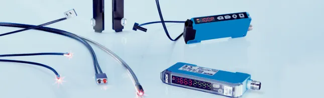
This is why newer monitoring approaches matter. Recent research describes systems that reconstruct broader temperature fields from limited sensor data, and reviews continue to emphasize that non-invasive or minimally invasive thermometry is central to making hyperthermia more usable, reproducible, and clinically safe. For applications where electromagnetic compatibility, flexibility, and multi-point sensing matter, fiber-optic approaches remain especially relevant.
What most teams get wrong about thermal safety
One common mistake is treating heat like a simple on or off variable.
It is not.
Thermal therapy safety depends on location, time, tissue response, and control. A treatment can be biologically weak even when it feels technically active. It can also become unnecessarily aggressive at boundaries that were never meant to receive high thermal exposure. Clinical reviews and current treatment guidance both keep coming back to the same principle: thermometry is not a side accessory. It is part of the treatment itself.
Where the field is moving next
The direction is clear. Safer thermal oncology will likely depend on better integration between planning, sensing, imaging, and feedback control. That means fewer blind spots, better spatial awareness, and more confidence that the achieved thermal dose resembles the intended one. It also means systems that can support deep tumors, challenging anatomy, and repeated treatment sessions without turning thermometry into an impractical burden.
Conclusion
In cancer thermal therapy, the safest teams are not the ones that simply deliver heat. They are the ones that can see, interpret, and control it. That is why temperature measurement in cancer thermal therapies is not just a technical detail. It is the bridge between intention and safety, between planned energy and actual tissue effect, and between a promising therapy and a dependable one. For engineering groups, device developers, research labs, and clinical teams that need support choosing or refining sensing strategies for demanding thermal environments, BioTemp4Life can help evaluate application fit, measurement approach, and next-step setup decisions.
FAQ
What makes a good temperature monitoring setup in thermal therapy?
A good setup measures the target and nearby risk tissue, supports real-time adjustment, and fits the anatomy without creating unnecessary workflow burden.
What are the best practices for safer thermal therapy monitoring?
Plan the risk zones first, use more than one meaningful measurement point, adjust during treatment, and review thermal dose afterward.
What trends are shaping thermal therapy monitoring?
The field is moving toward model-guided temperature mapping, real-time control, and better non-invasive or minimally invasive sensing.
How to choose between single-point and multi-point temperature measurement?
If the target is deep, irregular, or close to sensitive tissue, multi-point or mapped monitoring is usually the safer choice.
When to hire a specialist thermal sensing partner?
Bring in a specialist when anatomy is complex, heat distribution is hard to predict, or the treatment must work reliably across repeated sessions.
What services matter most from a specialist monitoring provider?
Application review, sensor selection, demo support, setup guidance, and help interpreting what the temperature data means in practice.


Sign in to leave a comment.